The significance of lower-extremity vascular assessment in patients with diabetes is both critical and often underestimated. Diabetes considerably increases the risk of vascular complications, especially in the lower limbs (Yachmaneni et al, 2023). Prioritising vascular assessments in patients with diabetes can support timely identification of vascular issues, which can lead to interventions that substantially enhance patient outcomes (Yachmaneni et al, 2023). By understanding a patient’s vascular health, healthcare providers can categorise individuals based on their risk of complications, enabling more personalised treatment strategies. Vascular assessments offer invaluable opportunities to inform patients about crucial foot care practices, necessary lifestyle modifications, and the importance of maintaining glycaemic control (Yachmaneni et al, 2023). As part of routine patient surveillance, follow-up assessments allow healthcare professionals to track disease progression and evaluate the effectiveness of implemented treatment plans (van Netten et al, 2024). Moreover, conducting a thorough diabetic foot assessment is crucial for identifying potential issues, particularly in the context of screening for limb perfusion to recognise early signs of peripheral artery disease (Bus et al, 2024).
Conducting routine vascular assessments allows healthcare providers to identify issues, such as peripheral artery disease, early, which is essential for preventing severe consequences, including ulcers, infections and potential amputations (Gul and Janzer, 2025). Addressing vascular issues early can significantly reduce the risk of more severe complications associated with diabetes (Gul and Janzer, 2025). In summary, lower-extremity vascular assessments are vital for effective diabetes management and can greatly enhance patients’ quality of life. Therefore, it is essential to integrate routine screenings as standard practice within diabetes care to achieve optimal health. However, the landscape of non-invasive vascular studies offers multiple methodologies by which to conduct vascular assessments, although none currently rise to the status of a definitive gold standard (Andersen, 2010).
Complicating matters is that standard non-invasive vascular studies can frequently produce misleading results in diabetic patients, which may unwittingly lead healthcare professionals to believe that adequate blood flow is present in the diabetic foot, especially in the context of diabetic peripheral neuropathy (DPN) (Dinh and Veves, 2005). Autonomic neuropathy can significantly contribute to the development of microvascular disease by impairing the regulation of blood flow due to nerve damage, thereby increasing the risk of serious complications, such as retinopathy, nephropathy and further neuropathy, in individuals with diabetes (Fowler, 2008). The autonomic nervous system controls involuntary bodily functions, such as blood pressure, heart rate and blood vessel dilation, which are crucial for maintaining proper blood flow throughout the body (Thomas, 2011). Damaged autonomic nerves disrupt the normal regulation of blood vessel diameter, causing issues, such as impaired vasodilation, which can lead to reduced blood flow to the tissues of the lower extremities resulting in functional ischaemia (Thomas, 2011).
The focus of this study was to explore and compare the microcirculatory responses of two distinct groups: individuals without diabetes and no symptoms of neuropathy and those suffering from diabetic neuropathy. By subjecting both groups to a carefully designed series of controlled stressors, the research aimed to uncover important physiological differences. A central goal was to clearly demonstrate that the microcirculatory stress response is markedly impaired in feet affected by diabetic neuropathy. Furthermore, the study sought to validate the efficacy of near-infrared spectroscopy (NIRS) as a reliable tool for detecting these significant changes in microcirculation, highlighting its potential role in enhancing clinical assessments for diabetic patients. Through this investigation, the study aimed to provide valuable insights that could lead to more effective management and treatment strategies for individuals living with diabetes.
Materials and Methods
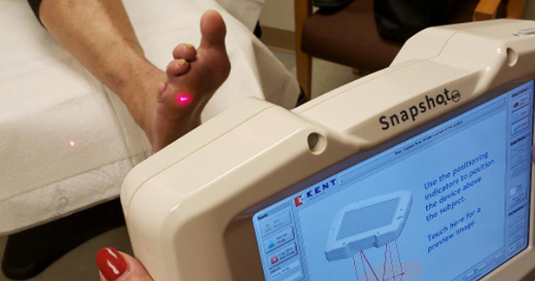
This prospective single-site case-controlled study compared the microcirculatory stress response in the diabetic and non-diabetic foot using near infrared imaging. This study received approval by Kent State University Institutional Review Board. The primary objective of this study was to demonstrate if the microcirculatory stress response is impaired in the diabetic neuropathic foot. The secondary objective was to demonstrate the clinical utility of NIRS (SnapShot NIR Kent Imaging, Calgary, AB, Canada) to detect changes within the microcirculatory system of the foot.
Cohorts consisted of people with diabetes and confirmed DPN and persons without diabetes. Patients with diabetes completed the two-page Michigan Neuropathy Screening Instrument and had a Semmes-Weinstein Monofilament test to verify the presence of DPN. Each cohort contained 10 patients. After a thorough informed consent process, each participant was screened using the study inclusion/exclusion criteria [Tables 1 and 2].
A total of 22 subjects were screened for participation in the study. Two subjects were excluded due to Ankle-Brachial Index readings that were outside the predefined inclusion criteria, thereby resulting in their classification as screen failures. Following the screening process, demographic data were collected from the remaining subjects, including age, gender, race, ethnicity, tobacco use, Fitzpatrick Skin Type Score and relevant medical history. Baseline NIRS images were captured from both the dorsum and plantar regions of the right foot while subjects were seated in a semi-recumbent position, prior to the administration of the targeted stresses. To maintain consistency throughout the study, the researchers designated the right foot as the index limb for all participants. This decision was implemented to ensure uniformity in the measurements and comparisons across the various subjects involved in the research.
Testing
Subjects had the right foot exposed to targeted stresses including in the following order: elevation, dependency, heat and cold for 5 minutes in a controlled environment. To minimise any carry over effect the subjects were returned to a semi-recumbent position for a rest period of 10 minutes between all stresses. NIRS imaging was obtained from the dorsal and plantar foot after completing each stress test.
Limb Elevation
The study limb was raised by a study team member standing at the foot of the treatment chair facing the subject. With the calcaneus gently cradled and a soft bend in the knee, the leg was elevated to a height to create 45 degrees ± 5-degree angle of the femur, as determined by the person performing the imaging when standing at the side of the bed/gurney. The limb was kept in this position for 5 minutes. Up to four NIRS images were taken of the dorsal and plantar surfaces of the right foot.
Limb dependency
The subject dangled the study limb over the edge of the treatment chair creating a 90-degree angle at the posterior knee bend. The limb was kept in this position for 5 minutes. Up to four NIRS images were taken of the dorsal and plantar surfaces of the right foot.
Heat
A 12”×24” electric heating pad on setting #5 (out of six) was wrapped around the right foot for 5 minutes. Up to four NIRS images were taken of the dorsal and plantar surfaces of the right foot.
Cold
A reusable cold therapy compress was wrapped around the right foot for 5 minutes. Up to four NIRS images were taken of the dorsal and plantar surfaces of the right foot.
Results
Subject Demographics
In this clinical trial, a total of 20 subjects were enrolled, with 10 participants assigned to each of the two study arms. The DPN group consisted of an equal distribution of gender, five males and five females. In contrast, the control arm included four males and six females. The demographic characteristics revealed a significant age difference between the two groups, with the mean age of the subjects in the DPN group being 64 years, while the control arm had a notably younger mean age of 37 years.
All participants in the DPN group identified as African American, highlighting a lack of racial diversity within this specific group. The control arm, however, presented a broader racial composition, including two African–Americans, two Caucasians, four Asians, and two Indians. It is important to acknowledge that issue oxygenation imaging has enhanced its applicability for patients with darker skin tones by incorporating melanin considerations for a significant number of individuals. Recent advancements in NIRS technology have improved the signal-to-noise ratio, thereby enabling better differentiation between melanin and both oxyhaemoglobin and deoxyhaemoglobin. This progress facilitates more accurate calculations of tissue oxygen saturation (StO2) in patients across the Fitzpatrick Skin Type Scale. Furthermore, the melanin correction process is automated, which minimises variability among a diverse patient population.
In terms of body composition, the mean BMI was markedly higher in the DPN group at 33.08, suggesting a prevalence of overweight or obesity, compared with the significantly lower mean BMI of 23.76 observed in the control arm, which indicates a healthier weight status among these individuals.
The Ankle-Brachial Index, a measure used to assess vascular health in the author’s clinical practice, revealed a mean value of 1.02 in the DPN group compared to a slightly lower mean of 0.98 in the control group, suggesting comparable vascular health between both groups.
Socioculturally, none of the subjects in the control arm reported any history of smoking, whereas the DPN group demonstrated a more varied smoking history: two individuals were identified as current smokers, two as former smokers and five reported no smoking history whatsoever.
Lastly, the mean score on the Modified Neuropathy Score Index, which offers insight into the severity of neuropathic symptoms, was determined to be 3.85 for the DPN group, indicating a moderate level of neuropathy among these patients. This comprehensive demographic, health and lifestyle profiling underscores the differences and potential implications of DPN compared with a control population [Table 3].
Dataset
Markers were positioned on both the dorsal and plantar images acquired via NIRS [Figure 2]. The StO2 values were recorded from the first toe (great toe), the fifth toe, and the midsection of the foot. Additionally, a fourth variable mean was computed by averaging the StO2 values from these three anatomical sites. Consequently, four StO2 values were recorded from both the dorsal and plantar aspects (two sides) of the NIRS images of the right foot of the subject, both at baseline and following four distinct stressors (elevated, dependent, heat, and cold) with a rest period of 10 minutes between stresses. This approach yielded a total of 40 variables for analysis, calculated as follows: four conditions multiplied by two sides multiplied by five measurement locations.
Figure 2 presents StO2 imaging of the dorsal (left image) and plantar (right image) surfaces of the right foot of a subject, as visualized using NIRS technology. The generated heat maps are designed such that regions depicted in blue or cooler tones indicate lower StO2 values, while those in red or warmer tones correspond to higher StO2 values. The values displayed within the markers located on the toes represent the mean StO2 of all tissues encompassed by each marker. Additionally, for the lasso drawn at the midpoint of the foot, both the mean StO2 value and the area of the lasso are provided in the accompanying black text boxes.
Four additional parameters were established by calculating the differences in StO2 values from the baseline (bs) position to elevated (ele), dependent (dep), post-heat treatment (heat), and post-cold treatment (cold) conditions. These differences are represented as bs_ele, bs_dep, bs_heat, and bs_cold, resulting in a total of 32 additional variables (calculated as four conditions multiplied by two measurements multiplied by four comparisons).
Statistical Analysis
All variables underwent analysis using a multivariate General Linear Model to assess statistically significant differences in values between the subjects with DPN and the control group. The results did not reveal any variables that significantly distinguished between the diabetic and non-diabetic groups. Nonetheless, some trends in the data were observed across the two patient cohorts, as depicted in Figures 3 and 4.
Observations
Figure 4 demonstrates that elevation results in a reduction of plantar StO2 values from baseline measurements for subjects in both groups. Furthermore, Figure 3 indicates that subjects with DPN exhibit a more pronounced decrease in StO2 values from baseline compared to subjects without DPN.
However, this phenomenon is not as distinctly observed on the dorsal side, as illustrated in Figure 3. Both Figures 3 and 4 convey that there is no discernible trend in StO2 values from baseline in relation to other stress factors, such as dependency, heat, or cold exposure.
Discussion
The findings presented in this study provide valuable insights into the impact of elevation on StO2 values in subjects with and without DPN. The observed decline in plantar StO2 values upon elevation suggests that alterations in foot positioning may significantly affect tissue oxygenation, particularly in individuals with DPN. This decrease might reflect underlying vascular impairments commonly associated with neuropathy, emphasising the importance of monitoring StO2 levels in clinical assessments and interventions for this population.
Notably, the dorsal side did not exhibit the same degree of reduction in StO2 values, indicating that the effects of elevation may be localized primarily to the plantar region. This discrepancy underscores the need for targeted evaluations in different anatomical areas when assessing oxygenation status in patients with peripheral neuropathies. Moreover, the lack of observable trends in StO2 values from baseline concerning other stressors, such as dependency, heat and cold, suggests that these factors do not significantly influence tissue oxygenation in this context. This finding may have implications for understanding the physiological responses to varying environmental conditions, providing a clearer picture of how external stressors impact StO2 levels in individuals with differing health statuses.
The pronounced differences in elevated StO2 values from baseline between subjects with and without DPN further highlight the necessity for tailored management strategies in those affected by this condition. By recognizing that individuals with DPN demonstrate greater susceptibility to changes in oxygenation, healthcare professionals can develop more effective treatment protocols aimed at improving vascular function and overall foot health.
Endothelial dysfunction and autonomic neuropathy are critical factors contributing to microcirculation issues in the patient with diabetic neuropathic foot (Veves et al, 1998). These factors can lead to functional ischaemia, even when blood flow seems adequate under normal circumstances (Dinh and Veves, 2005; Kolluru et al, 2012). NIRS has emerged as a validated technology, effectively measuring functional tissue oxygen saturation in diabetic foot ulcer management (Andersen, 2010; Neidrauer et al, 2010). The compelling results from this pilot study further support the idea that NIRS is not only instrumental in monitoring oxygen levels but also serves as a powerful tool for identifying changes in the microcirculatory system of the foot in patients with and without diabetes.
Limitations
The variables analysed in this study failed to show statistical significance in distinguishing between individuals with DPN and those without any health issues, primarily due to the constraints imposed by a limited sample size and lack of homogeneity. Specifically, there was a noted difference in age and weight between cohorts that could confound these finding. This restriction may have affected the reliability of the results and their ability to reveal meaningful differences between the two groups.
Conclusion
In conclusion, the findings obtained from this study significantly advance our comprehension of the relationship between elevation and StO2 levels, particularly concerning individuals diagnosed with diabetes who are experiencing neuropathic conditions. These results underscore the critical role of oxygenation in the overall health and wellbeing of this population.
Looking ahead, future research endeavours should aim to delve deeper into the intricate mechanisms that drive the observed changes in StO2 levels. Understanding these underlying processes will be crucial in developing targeted interventions that could alleviate the adverse effects of compromised oxygenation in individuals at risk.
Moreover, the trends noted in our data align closely with findings from previous studies employing NIRS technology in patients with peripheral artery disease (Andersen, 2010; Neidrauer et al, 2010; Reiter and Andersen, 2024). To strengthen the validity of our findings, it is recommended that this research protocol be expanded to include a larger and more diverse sample population, allowing for a more comprehensive understanding of these phenomena.