Da Vinci famously said: “The human foot is a masterpiece of engineering and a work of art.” With each foot containing 26 bones, four layers of intrinsic muscles, intricate neurological and vascular network and complex biomechanics, it is not difficult to see why this statement was made. But what about the frequently overlooked and underestimated lymphatic system? And how does this impact on the function of the foot and lower limb?
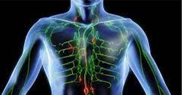
From an anatomical perspective, we know there are two main lymphatic paths from the lower leg: these being the fibular route to the popliteal node and the tibial route to the nodes in the groin. However, little is known about the lymphatics of the foot and especially the plantar surface — the sole. One small study using diagnostic imaging showed that some vessels start from the lateral edge of the plantar surface, some from the heel and just one each from the centre of the sole and the little toe. The imaging showed agglomeration to the tibial side and then in proximity (peri-adventitial) of the foot branch of the great saphenous vein (Uhara et al 2002).
So why should we think more about the lymphatics of the feet? For a start, everything that leaks out of the vascular system plus cell debris, microorganisms, inflammatory mediators and immune cells have to be reabsorbed initially via the lymphatic capillaries. Generally speaking, it is easy for the fluid and its contents to move due to hydrostatic and oncotic pressure gradients. But what is crucial and often forgotten is the fact that even more important is the external pressure applied to the fluids and the lymphatics via the contraction of the surrounding tissues when the foot is in motion. Sounds very positive doesn’t it!
A number of pathological conditions that commonly occur in the lower limb can result in disruption of the lymphatic system. An example of this is the management of individuals with diabetes-related foot ulceration (DFU). This condition places immense stress on the lymphatic system. In individuals with DFU, there is a level of immunocompromise, with underlying neurological and vascular changes, and commonly a level of bacterial colonisation and/or infection. This places additional load on the lymphatic system.
It is well understood that lymph flow is driven by intrinsic or extrinsic factors. In skeletal muscle, lymphatic function its driven by extrinsic factors that is the contraction/relaxation of the skeletal musculature. But what if there is atrophy of the muscles? We are aware diabetes-related motor neuropathy leads to atrophy of the intrinsic muscles of the foot. The skeletal muscles being replaced often by poorly defined fatty infiltrate. This leads to a change in foot shape and function, but also significant compromise with respect to lymphatic system function resulting in in immuno-compromised and infection prone, zones within the foot. Further, inflammatory responses are perpetuated due to the failure of the lymphatics to remove the range of cytokines from the affected tissues. Changes in joint range of movement and subsequent stiffness due to collagen crosslinking also have an influence.
When it comes to treatment of acute foot pathology, including tissue loss in DFU, it often involves immobilisation of the foot and ankle in knee-high irremovable devices to effectively offload the wound. This can be applied with or without compression. When the foot is immobilised or when there is substantial pressure exerted by the footwear which does not facilitate this process or if the muscles normally responsible for helping lymph to flow are dysfunctional or atrophied or totally useless and unable to contract or exert pressure changes?
We are aware that there is a limited range of tissue pressures over which extracellular fluid (and its contents) can enter the lymph capillaries and that surrounding tissue movement (generated by skeletal muscle contractions) helps this. But once the lymph arrives in the collecting lymphatics through while there are neurogenic and myogenic mechanisms helping lymph flow there is a need for active tissue pressure changes generated by skeletal muscle movements. Regarding this, when the walls of the collecting vessels are stretched it causes the pericytes to stretch which facilitates the propulsion of lymph from one lymph collector to the next until it enters a lymph node.
As best we know, the intrinsic lymphatic pumping mechanisms generate about two-thirds of the lymph flow with the remaining one third coming from the compression resulting from variations in skeletal muscle tone. So, foot and leg movement are critical in this clearance from these areas.
It has been reported in the literature that the presence of oedema in individuals with DFU is associated with poorer outcomes. A prospective study of 314 individuals with DFU demonstrated those with oedema were more likely to undergo amputation (Apelqvist et al 1990).
However, there is currently inadequate evidence to recommend compression in this population, with only three older studies (Akbari et al 2007; Armstrong and Nguyen, 2008; Mars et al 2008) that have evaluated the effectiveness of compression on wound healing and/or amputation, with all studies demonstrating a high risk of bias. Certainly, there is more important work to be done in this space.
There are a number of pertinent questions to ask … Could greater attention to managing the lymphatics in the feet provide the recipe for success for the management of acute and chronic injury and/or tissue loss? It is well described in the literature that compression therapy is effective part of the treatment of other wound types, however, foot wounds there is limited available evidence. What is the role of the podiatrist in this equation? Podiatrists are the foot and lower-limb experts — does this expertise extend to the lymphatic system? There is a desire within the profession to know more and be more involved, which is evidenced in the attendance of the lymph focused sessions at the recent national podiatry conference. Podiatrists can prescribe and apply compression therapy, but how often is this being completed?
Let’s now see what we can all do together to improve research-based information, knowledge and outcomes!

