- Skin tears can occur quickly, so prevention is important
As we age, skin naturally becomes thinner and less resilient towards force. Therefore, older adults are at higher risk of getting skin tears and more severe skin tears than when they were younger. Keeping the skin moisturised will maintain its elasticity and strength. Use fragrance free and hypoallergic products. Read the label if unsure, because many products contain preservatives that can be absorbed through the skin.
Implement a fall prevention programme, as many skin tears occur due to trauma during a near-fall, fall or attempts to lift the patient off the floor. With near falls and falls, patients can bump against objects (walls, corners) and acquire skin tears.
Have the patient wear clothing with long sleeves or use padded arm sleeves for greater protection. Applying leave-on products to moisten/protect dehydrated skin and applying moisturising lotion to damp skin improve skin elasticity and protection.
2. Skin tears are common in the elderly
The prevalence of skin tears has been studied in different contexts. In long-term care, it varies from 2.23 to 92%; in the community, from 4.5 to 19.5%; in acute cases from 6.2 to 11.1%; and in palliative care from 3.3 to 14.3% (Rayner et al, 2015). Reported incidence rates of skin tears ranged from 2.23% to 92% in long-term care facilities and varied from 2.1% among men to 4.6% among women living in the community.
3. There are many intrinsic and extrinsic risk factors for skin tears
Skin tears occur most often in the aged, especially those patients with thin skin, dry skin (cutaneous xerosis), who use corticosteroids, or have haematomas from a fall. Other intrinsic factors identified are being female, the presence of sensory, motor and balance deficits; altered mental state; malnutrition and dehydration; the presence of oedema; comorbidities. Among the extrinsic risk factors for skin tears is dependence on others for activities of daily living. Patients who require assistance for activities such as mobility, washing and dressing, are more at risk of skin tears due to manipulation and forces or trauma. Accidental falls are an additional extrinsic factor to consider, as these can cause skin trauma Other extrinsic risk factors are the use of adhesives, aids (orthoses/prostheses), wheelchairs, and feeding tubes (LeBlanc et al, 2011; Rayner et al, 2019; Cilluffo et al, 2023).
Skin tears can also occur during the removal of adhesives, such as tape and adherent dressings, which is also known as medical adhesive-related skin injury or MARSI (Cilluffo et al, 2023).
4. Avoid these actions to prevent skin tears
Hygiene with cold water and soap can increase skin tears. Having exposed arms by wearing short sleeves has been found to be associated with skin tears. Transferring patients into and out of bed in a rough manner and care providers wearing jewellery or long nails can increase the risk of skin tears. Devices with sharp edges in the resident’s room can lead to skin tears. Rapid removal of adhesive dressings or bandages can also cause skin tears (Cilluffo et al, 2023).
5. Control the bleeding first
Skin tears bleed profusely because of exposure to the papillary dermis, especially in patients who are anticoagulated, including those on aspirin. Common methods to control bleeding, such as pressure and elevation usually will need to be in place for up to 10 minutes. Excess dried blood and clots should be gently removed with normal saline. Syringes of saline work well for removal of dried blood. Do not scrap or scrub the skin to remove dried blood, as this can damage the blood supply to the skin tear and cause more skin tears.
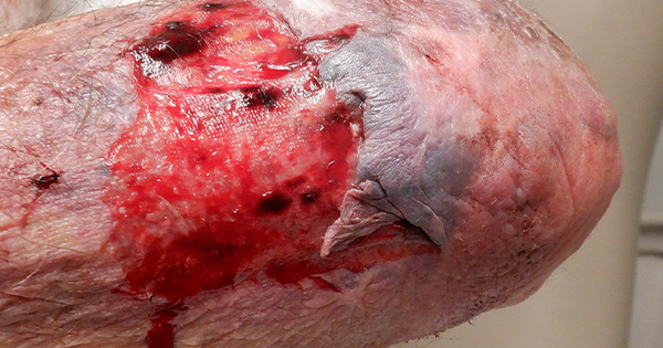
6. Classify the degree of injury using the skin tear classification system
The classification of skin tears has been described by the International Skin Tear Advisory Panel (LeBlanc et al, 2013). There are three types based upon the degree of skin loss [Figure 1].
Type 1: No skin loss, and the skin flap can be repositioned to cover the entire wound bed.
Type 2: Partial skin loss, and the skin flap cannot be repositioned to cover the entire wound bed.
Type 3: Total skin flap loss, exposing the entire wound bed.
7. Realign the skin flaps in type 1 and 2 skin tears
To realign the skin flaps, use a moist cotton swab. For a Type 1 skin tear without skin loss, reposition the edges to cover the wound bed. For a Type 2 skin tear with a partial flap loss, reapproximate the wound edges. Do not secure the skin flap with Steri-Strips because they are too difficult to remove. Skin sealants can be used.
8. Cover the wound bed with nonadhesive dressings for Type 3 skin tears
In a Type 3 skin tear with total flap loss and the wound bed entirely exposed, cover the entire area, preferably with a non-adhesive dressing. Wounds heal more quickly when the wound bed is kept moist and warm. Transparent films and hydrocolloid dressings are not recommended for skin tears due to their highly adhesive nature and inability to collect drainage. Hydrocolloids and transparent films can also lead to the development of skin tears (LeBlanc et al, 2011, 2013, 2018).
9. Indicate how the dressing should be removed
Mark the dressing with an arrow to indicate which direction the dressing should be pulled off for removal. The dressing should be pulled in a direction that is with the flap, not against the flap, to avoid lifting the flap.
10. Monitor for delays in healing
Skin tears can become infected, especially if on the leg or foot. Conditions such as diabetes, arterial disease, and immunodeficiency can delay healing and increase the risk of infection. Monitor for usual signs of infection including redness, swelling, warmth, pain, or purulent drainage.