When the concept of TIMERS (tissue, infection/inflammation, moisture, wound edge, repair/regeneration, social) was introduced in 2019, it outlined the components needed for hard-to-heal wounds (Atkin et al, 2019). If wound edges were not favourable, wound edge excision and evaluation for the possible need for therapies to accelerate re-epithelialisation were recommended. TIMERS also introduced the new concept of repair and regeneration, which focused on encouraging wound closure by providing a matrix to support cell infiltration, stimulating cell activity using signal molecules or growth factors, delivering oxygen therapy or using stem cells.
While repair and remodelling involve work with tissues, remodelling differs from repair. Repair focuses on fixing damaged areas to restore functionality. Remodelling involves making changes to the space and structure. Several new techniques were developed to fill the gaps, preserve wound edges, and remodel wounds with undermined areas and pockets to prevent their unroofing and resection.
The edge trenching and soap scrap techniques were based on the existing concept of fruit tree grafting, where one makes a fresh cut on the transplanted graft called scion (the upper part of the fruiting tree) and another cut on the rootstock (the bottom part of the accepting tree). The two are then bound together [Figure 1]. The wounded rootstock heals the new wound, and the scion engrafts. Translating this agricultural concept to wound care results in two walls of the wound being debrided and growing together under favourable circumstances (i.e. immobilisation and compression). The parallel pocket incision was developed to facilitate closure of pressure injuries with pockets. Even though soap scrap and parallel pocket incision techniques differ in methodologies, they have one common trait: a collapse and obliteration of the pocket cavity.
Below, we review four typical scenarios when wound remodelling is usually needed to achieve wound closure.
Wounds with raised base
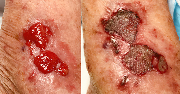
The wound bed needs to be at or slightly below the level of the periwound for the keratinocytes to cross the wound base from the periphery (Rousselle et al, 2019). Contouring the wound base to ensure it is not higher than the periwound can be achieved by debriding the base with a large-sized curette or razor blade (Melnychuk and Fox, 2023). If hypergranulation is present in a wound bed, chemical cautery with silver nitrate may be used. Debridement can also be combined with chemical cautery to speed up the process [Figure 2].
Wounds with vertical edges
Vertical edges are edges at right angles to the wound bed, which prevents smooth migration of keratinocytes from the edge to the centre of the wound.
One option for promoting healing in wounds with vertical edges is to resect the edges at 45° to ensure that the edge of the slope is smooth, and nothing impedes the keratinocytes from moving centrally. This approach works well for shallower wounds. If the wound is deep or the edge resection would create a significant tissue defect, the base can be raised first by using one of the following approaches:
Optimising tissue growth in the wound base (e.g. by applying collagen or extracellular matrix dressings, adding growth factors, etc).
Suturing a collagen scaffold (e.g. a bovine collagen dermal repair scaffold) to fill in the base of a deep wound [Figure 3]. After the collagen scaffold is placed, the wound may heal via primary closure or need to be followed by a split-thickness skin graft or myocutaneous flap.
Application of negative pressure wound therapy (NPWT) to promote granulation tissue in the wound base.
Using a combination of the above modalities (e.g. placement of collagen dressing under NPWT foam or the use of NPWT to bolster collagen scaffolds).
When the wound becomes shallower, the edges can be resected to 45°. When it is not possible to resect edges at 45° or if the walls of the wound do not merge with the base of the wound, the edge trenching technique can be attempted.
An edge trenching technique can be utilised when it is not possible to debride the wound edge to a 45° angle or when the wound base and the edges do not merge (Melnychuk, 2023). Often, these two clinical scenarios coexist. For this procedure, a small curette is used to excavate a trench at the point of convergence of the wound base and the wound wall [Figure 4]. The base of the wound and the edge wall are resected approximately at a ratio of half and half. As the newly created trench heals, both wounds contract and create a favourable transitional angle [Figure 5].
If wound edges change shape with body movement (e.g. if the wound is located over a flexible joint), it is important to immobilise the edges to allow the crosslinking process to take place uninterrupted. The procedure is repeated at each visit until the connection between the two wound surfaces is visible to the naked eye.
Wounds with rolled edges
Rolled edges form when wound contraction occurs while keratinocytes migrate from the edges and follow the wall. When the wound contracts, the edge creates an awning or overhang. The underside of this overhang gets fully epithelialised and subsequently does not adhere to the tissue in the wound base. Typically, it is recommended to resect these rolled edges or cauterise them with silver nitrate. When this is not possible or practical, the epithelial tissue on the underside of awning can be denuded with a curette. Then, pressure is applied using a compressive multilayered wrap, a combination of traditional and closed incision negative pressure therapy, or both. When the rolled edge is finally attached to the wound base, one can resect the edge at 45° or create a trench to help merge the base and the edge.
Wounds with undermined areas and pockets
Undermining is difficult to examine and, as a result, may be difficult to debride. Wound pockets are contained undermined areas. Typically, it is recommended to completely unroof and expose the undermined areas. However, there are two salvage approaches that can be attempted without resection of the undermined areas.
The soap scrap [Figure 6] technique consists of debriding contact wound surfaces, immobilising tissues in areas where tissues move, and utilising pressure (multilayered compression alone, a combination of conventional/closed incision NPWT, or both) to collapse undermining and wound pockets (Melnychuk and Juriga, 2023).
This approach follows the old thrifty practice of reusing two left-over soap bars. Both soap bars are softened (i.e. debrided), then compressed against each other (by applying NPWT, compression wrap or both) and finally, they are left alone for some time (immobilised) until they become one. This method works especially well on wounds located on the extremities; it is less successful when applied to lower back wounds, most likely due to increased depth or inability to immobilise tissues well.
The parallel pocket incision technique has been used in the treatment of pressure injuries with a pocket (Yamamoto et al, 2015). One incision is made at the proximal end of the pocket and another at the distal end, allowing thorough debridement of the pocket that is subsequently obliterated. [Figure 7].
No compression of the cavity is used in these cases, and the cavity is allowed to obliterate on its own. The authors have also successfully used a combination of parallel pocket incision and soap scrap techniques when treating extremity haematomas. To perform this, make two incisions at the proximal and distal end of a haematoma, drain it, mechanically debride any necrotic tissue in the cavity, and collapse the cavity using compression wraps. This approach may obliviate the need to unroof extremity haematomas.
Conclusion
Wound remodelling is a crucial concept that wound practitioners need to understand when encountering wounds with unfavourable topography of the wound base or edges. Wound remodelling may be part of TIMERS, where R would stand for repair, regeneration and remodelling. Wound remodelling techniques, such as edge trenching, soap scrap and parallel pocket incision, are tissue-preserving techniques that belong in the toolbox of every wound care practitioner. Additional operative techniques aimed at closing complicated wounds are needed.