Diabetes in South Africa is a major challenge for patients and the country itself, with the country having the second highest number of people living with type 2 diabetes in sub-Saharan Africa (Sifunda et al, 2023). The prevalence of diabetes has rapidly increased in South Africa, from 4.5% in 2010 to 12.7% in 2019. Of the 4.58 million people aged 20–79 years who were estimated to have diabetes in South Africa in 2019, 52.4% were undiagnosed (Sifunda et al, 2023). It is suggested that increasing household income and urbanisation has accelerated changes in environmental and social stressors, diet and physical activity behaviours of South Africans, predisposing them to increased risk of a range of non-communicable diseases, including diabetes (South Africa Demographic and Health Survey, 2016).
Diabetes-related wounds have been defined as a break of the epidermis and at least part of the dermis in a person with diabetes. The pathway to ulceration, involving loss of sensation, ischaemia and minor trauma (McDermott et al, 2023). The underlying aetiology of diabetes related ulcers is classified into three types: purely neuropathic (35%), purely ischaemic (15%) and mixed neuroischaemic (50%; Sidawy et al, 2018). Morbidity following diabetes-related ulceration is high, with recurrence rates of stated to be around 65% at 3–5 years, a lower-extremity amputation incidence of 20%, and 5-year mortality of 50 to 70%. Recent data state that overall amputation incidence has increased by as much as 50% in some regions, especially in young and racial and ethnic minority populations (McDermott et al, 2023).
Furthermore, in the South African public healthcare sector, 28% of patients living with diabetes present to primary healthcare clinics with diabetes-related wounds, often presenting in advanced stages of severity (Turner et al, 2024).
Wound infection
Wound infection presents clinicians with challenges, including higher resources and higher costs, possible increased hospitalisation, and interventions needed (Frykberg et al, 2015; International Wound Infection Institute [IWII], 2022). For patients living with wounds, infection is equally associated with poor healing outcomes, social isolation, malodour, increased pain and anxiety, and overall impaired quality of life (Upton and Upton, 2015; Woo et al, 2018). Furthermore, if wound infection is diagnosed tardily or managed inappropriately, it may have consequences, including spreading infection, amputation, sepsis and mortality in extreme cases (Pickwell et al, 2015; Ndosi et al, 2018).
There is noteworthy evidence that people living with diabetes are at a higher risk for increased wound infections, wound dehiscence, and pathological scarring, because factors such as nutritional status and glycaemic control also significantly influence diabetic wound outcomes (Dasari et al, 2021).
Topical antimicrobials have long been used in wound care for millennia to manage infection, and it is thought that the current usage is being driven in part by an increase in antibiotic resistance (Fletcher et al, 2020). Antimicrobial dressings have been developed to manage wounds at risk of or with clinical signs of infection and to prevent the extension of local infection to the surrounding tissue (Lützkendorf et al, 2022).
Moreover, the antimicrobial efficacy of silver ions against many species of Gram-negative and Gram-positive bacteria and their biofilms, which are most frequently responsible for wound infections, as well as certain fungi, yeast and viruses, has been supported by literature and guidelines (Braunwarth et al, 2020; Wounds UK, 2021; IWII, 2022).
TLC-Ag
Technology lipido colloid with silver dressings (UrgoTul Ag, Laboratoires Urgo, France, TLC-Ag) are supported by both in vitro and in vivo evidence (Lazareth et al, 2007; 2008; 2012; Bison et al, 2013; Desroche et al, 2016). The lipido-colloid particles gel in contact with wound exudate, maintaining a moist environment favourable to the promotion of wound healing (White et al, 2015), while the Ag+ ions provide anti-inflammatory properties and antimicrobial activity, leading to reduction of the microbial load (Bison et al, 2013; Desroche et al, 2016).
The efficacy of TLC-Ag in reducing clinical signs of local infection and promoting wound healing has been demonstrated in a randomised controlled trial (Lazareth, 2012). In a large observational study from Germany, 728 patients with wounds of various aetiologies and wound infection status were treated with the evaluated dressings in 39 centres for a mean duration of 26 ± 19 days (Lützkendorf et al, 2022). At the initial visit, patients presented with different wound infection statuses. Wound infection was established in 440 (60.4%) patients, based on direct indicators and/or clinical signs of infection; 183 (25.1%) patients presented with the first clinical signs of a wound infection (but not yet an established infection); 96 (13.2%) patients were at risk of wound infection (but with no clinical signs of infection); and nine (1.2%) patients had an unclearly documented wound infection status.
The cases
In view of this evidence base, the author aimed to evaluate this dressing, in conjunction with evidence-based multidisciplinary approach, in the management of a cohort of patients with complex wounds at his outpatient wound clinic.
A multidisciplinary team approach is the most efficient way to treat many chronic conditions (Xu et al, 2023). In this case series, providers sought to implement a multidisciplinary approach to manage their patients with diabetes and diabetes-related wounds.
In compliance with ethical guidelines, informed consent was obtained from all patients included in the case series. These consents were documented in the respective patients’ hospital records. The study focused on real-world clinical observations, and all patient-identifiable information has been anonymised in accordance with ethical publishing standards.
Case 1
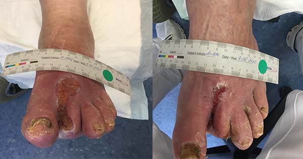
A 46-year-old woman living with poorly controlled, longstanding type 2 diabetes (T2D) and hypertension, presented on with ischaemic ulcers on three toes on her right foot [Figure 1A]. Her ankle–brachial pressure index (ABPI) reading was 1.3, indicating a false high, probably due to artery calcification. A CT angiogram was conducted, revealing early signs of intermittent peripheral arterial disease. Initial debridement was conducted to remove loose tissue and slough. A non-adherent technology lipido-colloid dressing with silver (UrgoTul Ag – TLC-Ag) was applied as the primary dressing to manage local infection, with a secondary layer of non-woven sodium carboxymethylcellulose (CMC) fibre dressing to manage exudate. The dressings were held in situ by means of simple crepe bandage. A “football dressing” offloading technique was applied (Radar and Berry, 2008). The patient was provided with education on walking with crutches, to help reduce pressure on the affected area. No systematic antibiotics were prescribed.
On day 4, after two dressing changes, the wounds were already showing progress, with slight wound area reduction and a healthier wound bed [Figure 1B]. The same management was continued and progression was noted [Figures 1C and 1D].
The wounds were considered healed by day 79 [Figure 1E], after 13 dressing changes with the TLC-Ag dressing.
These apparently simple wounds ere a challenge due to the patient’s longstanding poorly controlled diabetes and poor vascularity. The primary dressing of TLC-Ag was helpful in the early management of local wound infection and was continued throughout, as the wounds were considered to be at high risk of reinfection.
The holistic management of the patient included a multidisciplinary team consisting of podiatrist, medical officer and wound care nurse.
Case 2
A 46-year-old man with longstanding poorly controlled T2D, hypertension, and hyperlipidaemia and peripheral neuropathy. Patient presented with a right foot plantar ulcer that had been present for 8 months. The wound had previously been managed with a paraffin-impregnated gauze dressing, gauze and crepe bandage, without any progress. On presentation [Figure 2A], the wound was cleansed and debrided, TLC-Ag dressing applied as a primary dressing, a secondary layer of CMC fibre dressing and held in situ with a simple crepe bandage. The football dressing offloading technique was applied to reduce pressure in the area. No systematic antibiotics were prescribed.
After three dressing changes, the wound presented with a healthier wound bed and signs of epithelialisation on day 11 [Figure 2B]. and progress continued at day 22 [Figure 2C] after eight dressing changes). The wound was considered healed at day 25, a total of 13 dressing changes [Figure 2D].
The management of the wound with TLC-Ag combined with offloading and holistic management by the multidisciplinary team managed to resolve this wound within a relatively short period of time, considering that the wound was previously managed without any success for the previous 8 months.
Case 3
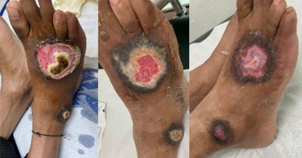
A 36-year-old man with longstanding T2D and hypertension, presented at the clinic 7 days post discharge for a metatarsal amputation. The wound was sloughy with unhealthy granulation tissue [Figure 3A]. Initial cleansing, desloughing and removal residual suturing was conducted. The treatment plan of TLC-Ag, CMC fibres and crepe bandage was applied for this patient. No systematic antibiotics were prescribed.
On day 11, after two dressing changes [Figure 3B], progress was seen, with less slough present. After a further two dressing changes [Figure 3C], wound progression was evident on day 25, with healthy granulation tissue and areas of epithelialisation. Visual evidence of further improvement was seen on day 57, after four further dressing changes [Figure 3D]. By day 103 (after five further dressing changes), the wound was considered healed and was covered with healthy epithelial tissue [Figure 3E].
In this challenging situation, the wound and the patient were managed by the multidisciplinary team. Although healing took 4 months, this case was resolved without having to resort to expensive and more challenging interventions, such as skin grafting.
Case 4
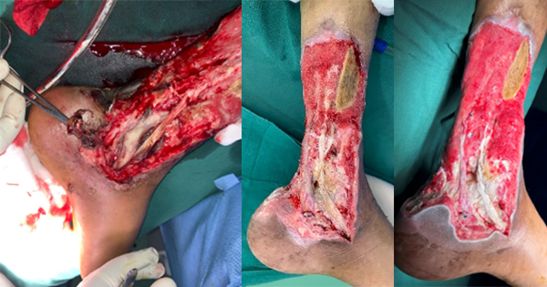
A 63-year-old man, living with uncontrolled T2D for many years, as well as hypertension, trauma-induced depression and osteoarthritis. He presented with a left foot Wagner Grade 2 neuropathic ulcer involving the tarsometatarsal area of the first, second and third toes, which eventually necessitated ray amputation of these toes (Shah et al, 2022). Postoperatively, the wound deteriorated and had he was referred 28 days following discharge from a surgical ward. Due to dressing shortages and lack of advanced wound care products at the primary healthcare clinic, he had been unable to receive adequate wound management.
On presentation [Figure 4A], the wound was highly exuding and malodorous, with a slimy slough covering all the wound. The wound was cleansed and initial desloughing conducted. The wound was again managed with the same treatment plan of TLC-Ag, CMC fibres and crepe bandaging. The football offloading technique was also utilised in this case.
By day 22, the wound bed was showing good signs of healthy granulation tissue, epithelialisation and reduction of exudate levels [Figure 4B]. Wound size reduction continued [Figures 4C, 4D and 4E]. At this point, the patient was referred to the primary healthcare clinic for further management.
This case presents another challenging wound due to the age and comorbidities of the patient. The multidisciplinary approach, holistic management and dressing regime proved to be effective even in this case in achieving positive outcomes.
Case 5
A 46-year-old man, with uncontrolled T2D, hypertension and hyperlipidaemia, presented with a 2-month-old neuropathic ulcer of unknown cause. Previously, the wound had been managed with povidone-iodine soaks, paraffin-impregnated gauze dressing, gauze and crepe bandage, without any improvement [Figure 5A]. Initial cleansing with hypochlorous acid solution and debridement were conducted, and the regimen of TLC-Ag, CMC fibres, crepe bandage and football offloading was applied, as well as provision of education on walking with crutches. After 1 month [Figure 5B], improvement in the wound bed was noted. After 2 months, further improvement and wound area reduction were evident [Figure 5C], and 2 weeks later, the wound had healed [Figure 5D]
This wound was previously managed in a ‘traditional manner’ which was not proving to be effective. Once again, the overall management by the multidisciplinary team and applying the TLC-Ag dressing as the primary layer, provided good results within the same time period of the previous traditional management.
Case 6
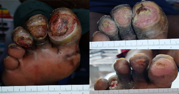
A 46-year-old man, with a history of uncontrolled T2D, hypertension, hyperlipidaemia, obesity and peripheral neuropathy, presented with a left foot plantar ulcer in the metatarsal area that had been persisting for 2 months [Figure 6A]. It was highly exuding with thick slough, and malodour, indicating possible infection. The wound edges were inflamed with epibole. The peri-wound skin was extremely macerated due to prolonged exposure to wound exudate. Debridement of slough and periwound skin was conducted and the TLC-Ag dressing and offloading was applied. Regular follow-up appointments were scheduled every week initially to monitor the wound. By week 4 [Figure 6B], the wound area was considerably reduced. Progress towards healing continued by week 8 [Figure 6C] and was completely healed by week 9 [Figure 6D].
Again, the involvement of the multidisciplinary team managed this case successfully, not only by implementing appropriate wound care, but also managing the patient holistically to ensure the best outcomes possible.
In all six cases, the dressing was rated a five (0 as the worst and 5 as the most desirable) in ease of application, ease of removal, non-adherence to wound, conformability, acceptability by clinician and acceptability by patient, without any adverse events reported.
Discussion
A document from Europe suggests that, although available evidence is limited, principles of antimicrobial stewardship should be adapted in the care of patients with wounds to reduce the unnecessary use of systemic or topical antibiotic therapy while ensuring the safest and most clinically effective therapy for infected wounds (Lipsky et al, 2016). This document recommends to initially apply silver dressings for a 2-week challenge period, after which the wound, patient and management approach should be re-evaluated to determine if a silver dressing remains appropriate. However, it should also be noted that some silver preparations have been shown as non-cytotoxic, and it has also been suggested that silver has actions that may promote healing (Ayello et al, 2012), while the TLC-Ag dressing has been reported to be safe over a period of 4 weeks (Lazareth et al, 2008).
From the author’s and his multidisciplinary team’s experience and point of view, they use antimicrobial dressings keeping in mind the environment and socio-economic risks of their patient population, especially those living in the rural areas. Nutrition, financial burden, socioeconomic and education status, and acute and chronic stress are variables that have either direct or indirect impact on wound healing (Fayne et al, 2020).
Conclusion
The positive outcomes achieved through the application of TCL-Ag dressing, based on the robust evidence of the TLC-Ag matrix, has provided the clinicians from Krugersdorp with a safe and effective option in the management of wounds with or at risk of local infection.