Lower limb lymphoedema is increasingly being recognised as a troubling symptom for people with advanced cancer. The literature suggests the incidence of lower limb lymphoedema in advanced cancer is currently between 25–80% (Landers and Thomson, 2017).
Lymphoedema occurs when there is compromise to the lymphatic system through a variety of mechanisms. Primary lymphoedema is the result of an abnormality in the lymphatic system. Secondary lymphoedema is from damage to the lymphatic system (Cooper, 2012). This latter is the type of lymphoedema commonly seen in advanced cancer.
Lower limb lymphoedema can cause a decrease in physical function and mobility, which can lead to a reduction in the quality of life for a person with advanced cancer (Bar-Sela et al, 2010). This can also place an increased burden of care upon the carers, in what is already a stressful time.
Discomfort, pain, infection, difficulty with micturition and psychological impact with physical appearance can all be distressing (Bar-Sela et al, 2010; Landers and Thomson, 2017).
This case study presents a novel approach to managing troublesome secondary lymphoedema in a patient with advanced metastatic cancer. Along with the improvement in the volume of lymphoedema, there was an unexpected psychological benefit for our patient. Together these improvements resulted in an improved quality of life.
Case presentation
Our patient was a 59-year-old man with metastatic anal squamous cell carcinoma, diagnosed one year prior. He had a large pelvic collection due to fistulising disease and was initially admitted to our inpatient palliative care unit for investigations into possible osteomyelitis and pain management. His lower limb lymphoedema and scrotal swelling became apparent 2 weeks into the admission. He was classified as ISL (International Society of Lymphology lymphoedema staging) stage 2.
Our patient was particularly troubled by not being able to wear his favourite boots and during his admission he gradually became unable to wear any of his footwear. This was problematic as he liked to walk out in the garden. The reduction in mobility and subsequent requirements for utilisation of a mobility aide was distressing for the patient; in addition to increasing pain and pressure in the leg that was refractory to analgesia.
The concept of subcutaneous drainage for our patient’s lymphoedema was discussed within the palliative medicine department.
With a limited prognosis, no appropriate medication options and many factors impacting on quality of life, there was a consensus to consider subcutaneous drainage. Magnetic resonance imaging (MRI) was consistent with tumour obstruction of the lymphatic system and our patient was hypoalbuminaemic (albumin19 g/l).
The procedure was discussed and consented to by our patient.
Previous case studies had reported using subcutaneous needle placement (Clein and Pugachev, 2004).
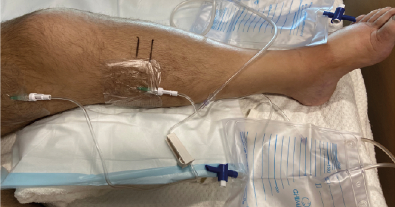
The decision was made to use subcutaneous cannulas to minimise pain. A closed drainage system was devised from equipment available (Figure 1). This drainage system consisted of an 18 gauge cannula connected to a catheter bag via standard tubing.
The left lower limb was more oedematous than the right; therefore, it was decided to try this procedure on the left leg only.
His initial weight was 88 kg. The left lower thigh circumference was marked and measured to be 54.0 cm and the left upper calf circumference was marked and measured as 47.6 cm. An application of 5% lidocaine cream was applied to the left medial thigh and left medial calf for 60 minutes.
Under sterile technique, two 18-gauge cannulas were inserted. The cannulas were attached to the closed drainage system (Figure 2) and secured with Opsite film dressings (Smith & Nephew) and crepe bandages.
Manual lymphatic drainage (MLD) massage was performed for the first 3 days to aid drainage. Regular circumference measurements were taken to monitor clinical improvement.
Two days after commencement of drainage, the patient reported that his shoe was fitting better. He was seen by the physiotherapist and able to mobilise 40 m with his walking frame. The MLD massages were providing important psychological benefit for our patient.
Medical review occurred daily to ensure that there were no signs of cellulitis. There was still a significant amount of oedema distally to the location of the two cannulas.
Our patient consented to further subcutaneous drainage. Following the same procedure, an 18-gauge cannula and a 22-gauge butterfly needle were inserted into the left leg, just proximal to the ankle. They were connected to the same type of closed drainage system. 24 hours later the butterfly needle had become displaced. The 18-gauge cannula just proximal to the ankle remained in situ and improved drainage efficiency.
On day 8, the cannulas fell out while the patient was in bed. They were not re-inserted as a positive result had been achieved for our patient. A total of 4.5 kg of weight loss had been achieved. The thigh circumference had reduced by 6.5 cm (from 54 cm to 47.5 cm) and the calf circumference had remained stable (48 cm).
Subjectively, our patient reported an improvement in mobility and comfort. He spoke with great positivity about the subcutaneous drainage. Later in his admission, he requested additional cannula insertion for lymphoedema re-accumulation.
Our patient died 5 weeks after the initial subcutaneous cannula drainage.
DiscussionSecondary lymphoedema
Secondary lymphoedema of the lower limb is commonly due to obstructive pathology or hypoalbuminaemia. Both were present in our patient. Extracellular fluids and their contents normally enter the lymphatic system throughout the limb, move upwards through the groin, abdominal and thoracic areas and empty into the venous system at or near the left jugular/subclavian vein junction. As a result, tissue pressure variations occur through movement and colloid osmotic pressures. Lymph passes through lymph nodes for purification and resolution of antigen presentation prior to being returned to the heart (Waugh and Grant, 2014).
Inability of the lymph to progress through the lymphatic system results in lymphoedema distal to the level of the damaged system.
Risk factors for secondary lymphoedema include surgery, radiotherapy, tumour progression, deep vein thrombosis, hyopalbuminaemia, renal or cardiac failure and medications (Cooper, 2012; Landers and Thomson, 2017). When lower limb lymphoedema occurs in advanced cancer, the common contributors are obstruction of the lymphatic system due to increasing tumour burden and hypoalbuminaemia. As lower limb lymphoedema progressively worsens, there is risk of breakdown in skin integrity and lymphorrhoea (Towers et al, 2010). This further increases the infection risk and makes mobility and care provision more challenging.
Current treatment options
The current gold standard treatment for lymphoedema is combined decongestive therapy (CDT) and MLD. CDT involves intensive multi-layer compression bandaging with a level of pressure that may not be tolerated in people with advanced cancer. The goal of standard CDT is to reduce swelling and allow for lifelong management of lymphoedema (Towers et al, 2010). This treatment goal is not as critical in advanced cancer. In a palliative setting, the goal of lymphoedema management is aimed at providing comfort and symptom relief, and maintaining the current level of function (Towers et al, 2010).
Subcutaneous drainage of lymphoedema
Subcutaneous needle drainage of lymphoedema in the advanced cancer setting was first described by Clein and Pugachev (2004). They reported on eight cases where they used subcutaneous needles and a closed system to allow for lymphoedema drainage.
Further case reports have been published about this method of lymphoedema drainage. Landers and Holyoake (2022) published a multicentre trial of subcutaneous needle drainage in a closed system design in 27 patients with an average drainage volume of 5.5 l. They also showed an improvement in quality-of-life markers.
Bar-Sela et al (2010) published a case report on eight patients with a combination of closed and open system setups. The closed system was the same as previous case reports used. The open system involved a subcutaneous tract being formed but with the needle removed at that time and open drainage occurred. Lymphoedema fluid loss was demonstrated with both types of systems.
Currently there is no determined gold standard method for this procedure.
In a palliative setting, goals of care must always be patient centred and realistic for the situation. The goals for this patient were successfully met because he had improved physical function, decreased pain and pressure, and a subjective improvement in his mental state following successful subcutaneous cannula drainage. Our patient had similar characteristics to other patients in the literature. He had advanced metastatic cancer with the lymphoedema having significant impact on his comfort and physical function. He also had a guarded life expectancy.
Previous case reports and studies have shown similar improvement in these important parameters of quality of life in advanced cancer.
The amount of weight loss experienced by our patient is consistent with other case reports (Bar-Sela et al, 2010; Cooper, 2012). Landers and Holyoake (2022) reported similar improvements in quality of life based on the Lymphoedema Quality of Life tool.
Our procedure differed by using cannulas rather than needles for the drainage process. Other case reports and studies have used needles only (Clein and Pugachev, 2004; Bar-Sela et al, 2010; Landers and Thomson, 2017; Landers and Holyoake, 2022).
Pain with the placement of needles has not been specifically mentioned in the case reports.
Consideration for patient movement with a needle in situ will need to be explored further when determining standardised procedures. Initially, there were concerns that the pressure from the lymphoedema would cause the cannula to collapse. However, the cannula remained patent and allowed drainage while in situ. The cannula also allowed a tract to form that allowed for ongoing lymphorrhoea once the cannula was removed.
Currently, no superior method has been identified to optimise lymphoedema drainage. Standardisation of this procedure would allow it to become more widely accepted as a valid treatment option in secondary lymphoedema due to advanced cancer.
Using a cannula may also allow this procedure to occur in the home setting and not necessitate an inpatient admission which would be in keeping with some palliative patient’s wishes.
Additional benefits
Another important aspect to this case was the subjective psychological improvement in our patient.
Cicely Saunders, the founder of the first modern hospice, has previously said: “The way care is given can reach the most hidden places and give space for unexpected development” (Saunders, 1996).
Our patient received MLD from one of the hospice nurses trained specifically in lymphoedema management. MLD not only potentially improved the drainage but also subsequently provided an opportunity for therapeutic touch and conversation which provided significant psycho-social support to a patient who has otherwise led a reclusive and isolated life.
The significance of improving psychological wellbeing in a patient with advanced metastatic disease should never be underestimated, although it is difficult to measure objectively.
Subcutaneous drainage of lower limb lymphoedema is a relatively simple procedure that provides a significant impact on a person in the last few weeks of their life. There will always be risks of infection in an immunocompromised patient; however, the benefits of subcutaneous cannula drainage extend beyond simple fluid loss.
Quality of life factors, including appearance, symptoms of pain and pressure and psychological improvement, have all been shown to improve following reduction in lymphoedema burden (Clein and Pugachev, 2004). Symptomatic improvement and psychological wellbeing can have a greater impact on a person at the end of life. The benefits appear to outweigh the risks in optimising quality of life for a patient with advanced cancer.
Conclusion
Subcutaneous drainage of lymphoedema with wide bore cannulas was successful in this case. Despite concerns about hydrostatic pressure, the cannulas remained patent, allowing for a 5% reduction in the patient’s body weight.
Future research in this challenging population group should focus on standardising the procedure for subcutaneous drainage and determining the most appropriate locations for cannula insertion to optimise drainage volumes. In advanced cancer, with a heavy lymphoedema symptom burden, being able to improve mobility and reduce the pain and pressure from lower limb lymphoedema will go a long way in enhancing quality of life for these patients.