A burn wound occurs when the skin comes into contact with a heat source, such as fire/flame, hot objects, electrical appliances and chemical agents. Injuries related to a burn are highly variable in their severity (Toussaint and Singer, 2014), where morbidity and mortality tend to increase in accordance with the surface area of the burn increase (Vivó et al, 2016). The causal agent, temperature, duration of contact, the location of the wound and its extended area, as well as the timing for its presentation to a healthcare professional are known to be contributing factors to the severity of the burn (Evers et al, 2010).
Burns have been defined as a global public health problem by the World Health Organization (WHO), accounting for an estimated 180,000 deaths annually. The vast majority of deaths occur in low- and middle-income countries and almost two thirds occur in the WHO African and South-East Asia regions. Non-fatal burn injuries are also a leading cause of morbidity (WHO, 2023). According to the National Programme for Prevention and Management of Trauma and Burn Injuries (NPPMTBI) from India, over 1 million people are moderately or severely burnt every year in the country, with an estimated 700,000 hospital admissions annually (Newberry et al, 2019). The high incidence has been attributed to illiteracy, poverty, and low-level safety consciousness in the population (NPPMTBI, 2023).
The priorities of specialised facilities and wound care clinicians dealing with burn wounds are to focus on stabilising the patient, prevent infection and optimise functional recovery (Rowan et al, 2015).
Burn infection
Sepsis continues to be the primary reason for death in burn patients (Norbury et al, 2016). Generally, microorganisms will colonise and grow quickly after burns (Mayhall, 2012), and infection of burn injuries is considered a common complication (Naqvi et al, 2014).
Burn trauma destroys the first line of defence provided by the skin and contributes to the suppression of the immune system, while the skin debris, and the protein-rich eschar that sometimes forms, offer an ideal environment for microorganisms to grow (Valarmathi et al, 2013; Dahag et al, 2018). Burn wound infections have been shown to be associated with wound deterioration, skin graft failure, hospital admission and/or prolonged hospitalisation (Cartotto, 2017). Pathological scarring in burn wounds can result in hypertrophic scars and/or contractures (van Baar, 2020).
Notwithstanding burn depth and extension, topical antimicrobials are indicated when there is clinical suspicion of risk of infection, or when wound infection is evident (Cartotto, 2017). The choice of local treatment is based on factors such as wound depth, anticipated time to healing, need for surgical intervention, known cytotoxicity of the agent, pain or irritation, and the associated frequency of application required (Cartotto, 2017). Advances in antimicrobial therapies have certainly added to the armamentarium of resources for the clinician (Norbury et al, 2016).
Local wound therapy
Burn wounds are prone to rapid bacterial colonisation with the potential for invasive infection and, therefore, measures to reduce the likelihood of infection are necessary (Tenehaus and Hans-Oliver, 2018). In India, standard of local treatment of burn wounds includes cleansing, debridement and routine burn wound dressing changes, often incorporating a topical antimicrobial agent (Tenehaus and Hans-Oliver, 2018).
Antimicrobial dressings are applied topically to the wound, providing a broad spectrum of non-selective antimicrobial action (International Wound Infection Institute (IWII), 2022). It is important, when considering wound antiseptics, to select agents that provide immediate onset and long-lasting broad-spectrum activity towards microorganisms but also a safe activity on both healthy and injured skin, as well as providing good tolerance without adverse effects (IWII, 2022)
The improvement of effective topical antimicrobial agents for burn wound care was possibly the single most important advance in the management and prevention of infection in these cases (Cancio, 2021). Traditionally, burn wound infection had been managed with silver sulfadiazine (SSD) cream (Cancio, 2021). However, although SSD had been considered the gold standard therapy in this indication for many years, its negative impact on the wound healing process has also been well documented (Abul Barkat et al, 2023), reporting inferior outcomes when compared with other antimicrobial agents (Genuino et al, 2014; Rashaan et al, 2014).
In a study conducted by Rosen et al (2015), several undesirable outcomes were associated with the use of the SSD cream, including notably a significant wound healing delay due to the cytotoxicity of the agent, but also to the formation of a persistent crust overlying the wound bed and slowing down re-epithelialisation. This yellow-grey pseudo-eschar crust, that can be several millimetres thick, seems to result from the direct interaction between the cream and the wound exudate (Bessey, 2007). Cytokine analysis on day three also showed the absence of several proinflammatory factors, and the authors suggested that the application of SSD might alter gene expression of IL-1-family genes, leading to impaired downstream cytokine production and ultimately delayed wound healing. Finally, it has also been suggested that SSD is relatively short-acting, time-consuming and messy to apply and remove (International Consensus, 2012), and that the lipid base of the cream make its removal painful for the patient (Black and Drake, 2015).
Technology lipido-colloid with silver sulfate (TLC-Ag)
Technology Lipido-Colloid (TLC), developed by Laboratoires URGO (France), is a matrix containing hydrocolloid and lipophilic substances. The TLC matrix promotes fibroblast proliferation through the creation of a moist wound environment, which in turn also contributes to the formation of new tissue (Bernard et al, 2005; McGrath et al, 2014). Moreover, it has been shown to reduce adherence to the wound surface (Meaume et al, 2002), therefore, allowing atraumatic removal and pain-free dressing changes (Meaume et al, 2004).
With the incorporation of silver sulfate (3.5%) to the TLC healing matrix, the TLC-Ag dressing (UrgoTul Ag/Silver® – Laboratoires URGO, Chenove, France) provides antimicrobial properties and is indicated for the local treatment of wounds at risk or with signs of local infection: chronic wounds (pressure ulcers and leg ulcers) and acute wounds (partial-thickness burns, dermabrasions, traumatic wounds and surgical wounds). It can also be used on more heavily exuding wounds when used in combination with an absorbent dressing (White et al, 2015). The antimicrobial efficacy of the dressing was first assessed in an in vitro study that showed that, from the first hours and throughout the duration of the study (7 days), the reduction in the number of colony forming units for all the bacterial strains studied, including MRSA and VRE, was >105, with 99.99% efficacy in just 24 hours (White et al, 2015). The TLC-Ag was reported as having the ability to destroy Staphylococcus aureus and Pseudomonas aeruginosa biofilms and to have antimicrobial effects on numerous organisms (Quatravaux et al, 2008; White et al, 2011) and in vivo to have significant anti-inflammatory effects on chronic skin inflammation (Bisson et al, 2013).
The clinical efficacy of the dressing has been demonstrated in a multicentre, phase III, controlled, randomised trial where the first arm of the cohort was treated for 4 weeks with the TLC-Ag interface dressing then for four weeks with a neutral TLC dressing, while the second arm of cohort (the control group) was treated with the same neutral dressing for 8 weeks (Lazareth et al, 2012). The results showed that the clinical score for wound colonisation, as defined by the presence of clinical signs among five pre-specified signs at baseline, was significantly more reduced in the TLC-Ag dressing group than in the neutral dressing group (1.43 versus 2.31, respectively [P=0.0001]). Moreover, a superior surface area reduction was achieved in the TLC-Ag sequential group than in the control group (median decrease at week 8 = 5.9cm2 vs 0.8cm2; P=0.002), presenting evidence that the dressing significantly promotes the wound healing process, in addition to providing effective antimicrobial activity.
Furthermore, the efficacy of the TLC-Ag dressing was evaluated in the management of almost 5,000 patients with chronic wounds including venous leg ulcers, arterial leg ulcers, pressure ulcers, and diabetic foot ulcers, as well as acute wounds, including traumatic and postoperative wounds in a prospective observational trial conducted in Germany and France. The results portrayed a healing rate of nearly 30% in less than 6 weeks, and also the progress of a steady granulation tissue, the substantial improvement of the clinical signs of infection. Moreover, Lützkendorf et al (2022) conducted a large multicentre observational study, including 728 patients with wounds of various aetiologies and wound infection status treated with the TLC-Ag dressings for a mean duration of 26 days, with an intermediate visit conducted in 97.8% of patients after a mean period of 12 days. Among these patients, 59 had a burn at risk of or with clinical signs of local infection. These burns were located on the lower or upper limbs (50.8%); on the hand, fingers, foot or toes (33.9%), on the abdomen or thorax (11.9% ) and at other locations (3.4%). The mean total body burn area was 3.8±4.3% (ranging from 1–18%), and the median wound area treated with the TLC-Ag dressing was 16.5cm² (IQR: 6.3–28.3cm2). A burn complication was also reported in 42.4% of the patients: with 12 cases involving large joints, nine cases circumferential extension and four cases periorifice location. Throughout the study period, all the parameters of wound infection continuously decreased in the total cohort, resulting at the final visit in a reduction by 78.9% of the prevalence of local wound infections and by 72.0% of the clinical signs of wound infection, the most rapidly diminished clinical sign being wound deterioration. Concurrently, in terms of the healing process, 92.1% of the wounds healed or improved, 3.2% remained unchanged and 1.7% worsened (data missing for 3.0%), and an improvement of the periwound skin was reported in 65.7% of the patients. Overall, the dressings were ‘very well accepted’ by the majority of patients, with no uncomfortable feeling at wearing and no pain at dressing removal. The dressings were assessed by the physicians as ‘very useful’ in the majority of the cases with a ‘very good’ efficacy in terms of antimicrobial activity and promotion of the wound healing process. Similar results were reported regardless of the wound type treated or of the TLC-Ag dressing evaluated or the wound infection status at baseline.
In India, another interesting case series presented five burn cases that were managed by the TLC-Ag dressing (Uppal et al, 2020). The authors concluded that managing burn patients in India with the TLC-Ag showed promising results, both in wound healing progression, but also in reducing wound pain and avoiding procedural pain. The same dressing was also evaluated with good results in different wounds by a group of clinicians from China (Yingtao et al, 2021). Interestingly, in a case series by Qiuju et al (2020), an infant with congenital skin deficiency was initially managed with TLC-Ag. What is interesting is that the child was suffering from G-6-PD deficiency, where sliver sulfadiazine is not recommended as it could be absorbed through burn and trigger haemolysis in G6PD deficient individuals (Albagshi et al, 2021). In view of the above evidence, the authors decided to evaluate the performance of the TLC-Ag dressing in their burn patients, which included two adults, one infant and one child. All the patients or legal guardians included provided informed consent to the use of the cases for publication.
Case 1 — Dr Venkateswaran Plastic Surgeon. Jupiter Hospital, Mumbai, India
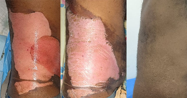
A 32-year-old male patient, labourer, was referred with scald burns on the left lumbar and iliac region, and anterior elbow region [Figure 1a] due to an accident at his workplace. The wounds were debrided and cleansed with sterile water and TLC-Ag was applied as a primary dressing [Figure 1b; 1c]. The dressings were changed on the fifth day, with substantial healing noted [Figure 1d; 1e]. There was a significant reduction of wound surface area after the application of TLC-Ag, with healing achieved after just one dressing change. No pain associated with the dressing was reported.
Case 2 — Dr Ravichander Rao A Plastic Surgeon. Care Hospital, Hyderabad, India
A 4-day-old male infant was brought to the hospital after sustaining an accidental scald burn from spilled hot water. The burn was located on the left foot, extended from the toes to the heel and ankle [Figure 2a]. The skin had peeled off on the entire foot sole, the heel and of some part of the first and fifth toes. The wound was debrided and cleansed with sterile water and a TLC-Ag dressing was applied as a primary layer, with dressing changes every alternate day. After the first dressing change, wound healing was progressing positively [Figure 2b]. Complete epithelialisation was achieved after 8 days and two dressing changes [Figure 2c]. In this very young infant case, healing with TLC-Ag was noted to be very rapid, and stress-free due to the absence of pain at dressing removal (no reaction from the child when the dressing was removed). Dressing application was reported as easy due to the flexibility of the dressing.
Case 3 — Dr Krishna Kumar,Plastic, Aesthetics, Burns Hand and Reconstructive Microsurgeon, Kovai Medical Centre & Hospital, Coimbatore, India
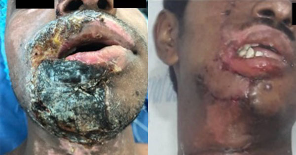
A 45-year-old male patient, working as a solar water heater technician, presented immediately after sustaining a right lateral side burn due to a solar water heater blast [Figure 3a]. The large wound was debrided and cleansed with sterile water and a TLC-Ag dressing was applied as a primary layer. The dressing was changed after three days; on the fifth day wound healing progress was already observed [Figure 3b]. Thereafter, dressing changes were scheduled less frequently. At the third dressing change, after 14 days of treatment, the wound was fully epithelialised [Figure 3c]. At the follow-up visit, 6 weeks after the burn injury, the aesthetic appearance of the healed skin was very good, including in term of skin coloration [Figure 3d]. It was noted that application of the TLC-Ag dressing resulted in a significant and rapid reduction of the wound surface area, with healing obtained after only three dressing changes without any complications. Pain score was rapidly reduced and, moreover, there was no associated procedural pain.
Case 4- Dr Sankamithra Consultant Plastic Surgeon, Lakshmi Medical center, Pollachi, India
A 4-year-old male boy presented with scald burns on the dorsal [Figure 4a] and ventral [Figure 4b] regions of his left arm and hand after he put his hand in boiling sambar (South Indian stew), 24 hours after the incident. Early excision of the burn regions, down to the fascia, was performed under anaesthesia in the operating theatre. The child developed compartment syndrome and, hence, fasciotomy was done to establish blood flow [Figure 4c; 4d]. TLC-Ag was then applied as a primary dressing, with dressing changes every 4 days [Figure 4e; 4f]. The wounds were healed after 8 days and two dressing changes [Figure 4g; 4h]. It was noted that there was a rapid significant reduction of wound surface area and pain score, as well as no associated procedural pain. Finger and hand functions were restored with good healing.
Discussion
Burn wounds are challenging in many aspects, more so because the injuries entail a vulnerability to colonisation by invading microorganisms and deficiency of the immunosuppressive system (Tsolakidis et al, 2022). Especially when wound closure is delayed, multi-resistant strains can develop and lead to serious complications (Trupkovic et al, 2012).
A diverse range of topical antimicrobial agents have been developed over the past decades. More recently, more modern interventions and utilisation of topical therapies have emerged (Ozhathil and Wolf, 2022), where topical antimicrobials have been associated with a decline in the incidence of sepsis due to burn wound infections (Lachiewicz et al, 2017). Antimicrobial dressings are arising as a promising approach for microbial burden control and wound healing (Yousefian et al, 2023). Silver is recognised for its clinical efficacy as a topical antimicrobial, and, the introduction of silver-impregnated dressings has increased its utility in the field to encompass a broader range of wound types, whether wounds are colonized or infected (Grada et al, 2019).
The evidence behind the efficacy of TLC-Ag directed the authors to evaluate the dressing in burn patients of different age groups. It has been previously stated that ‘the use of silver dressings in local wound care for children or infants is possible in the individual case on the basis of medical judgement and under medical supervision’ (Böttrich et al, 2019). The previous clinical evidence was (deemed) strong enough to support the safety of TLC-Ag in the case of an infant and young child.
Decreasing pain and distress experienced during a dressing change can have positive implications psychosocially for the children and their caregivers, as well as encourage re-epithelialisation of the wound within the optimal healing timeframe (Gee Kee et al, 2013). Therefore, the atraumatic properties of the TLC-Ag dressing was another aspect considered as optimal for this evaluation. Moreover, the highly satisfactory outcome in terms of skin aesthetics, notably in patients with dark skin, should also be remarked.
The results of these four cases are very satisfactory, prompting further evaluation of TLC-Ag to confirm the inclusion of this dressing as part of the standard of care for burn wound management in the different facilities.
Conflict of interest
All authors have no particular conflicts of interest to declare regarding these cases.
Limitations
The management of these patients was initiated based on positive outcomes obtained by previously published clinical evidence. The authors acknowledge that portraying only four cases may not be enough to standardise burn management in their facilities, but owing to other available evidence, in conjunction with these positive results, the inclusion of the dressing in further protocols might be considered and likely.