1. Be certain that the staff caring for the patient understand what should be done when problems develop.
While most NPWT systems work well, the drape can loosen and causes the suction to be lost. If the drape is loose, it can be reinforced with more adhesive dressings. If the power to the device is lost, the NPWT dressings must be removed and replaced with moistened gauze. The loss of power allows the drainage in the foam and tubing to drop back onto the wound. If the drainage cannister contains bright red blood, the NPWT system should be removed and the bleeding stopped with pressure.
2. Be aware of known contraindications to NPWT use.
Since the inception of NPWT it has been contraindicated on malignant tissues because the changes in the cell wall could increase tumor growth. Examine the wound for any exposed organs or blood vessels with each dressing change, do not reapply NPWT because bleeding or organ damage could occur. NPWT is also contraindicated with untreated osteomyelitis as a primary treatment due to the risk of promoting bacterial growth from the wound dressing remaining on the infected bone.
3. Be certain the wound has been adequately debrided prior to use.
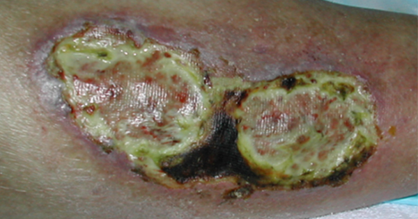
NPWT removes exudate, reduces oedema, reduces bacterial load, promotes blood flow and promotes granulation tissue formation (Ying Zeng et al, 2026). However, NPWT cannot replace debridement. Virulent bacteria, especially anaerobes, can multiply and spread if the wound is sealed without proper cleaning. If the wound contains dry necrotic eschar, slough or other non-viable tissue, the negative pressure may not be effectively delivered, and the dead tissue is a nidus for infection. Enzymatic agents on slough under the NPWT foam will have little effect on slough because the negative pressure will remove them from the wound bed (Bradley, 2014). Consider NPWT systems with instillation options to treat wound infections, but they are not a substitute for debridement [Figure 1].
4. Cleanse the wound before applying NWPT on subsequent dressing changes.
Wounds treated with NPWT can develop an odour, which is usually from the occlusive nature of the treatment and bacteria on the wound surface. The wound bed must be cleansed to remove debris, cells and bacteria. Antiseptic agents are preferred. Consider both the antiseptic properties and the cytotoxic properties of the agents used. Simply drizzling the antiseptic over the wound bed is not adequate, use a wound cleaning device and rinse the wound bed well.
Further, remove any newly formed slough in the wound bed. Slough should be removable with either a curette, gauze or wound cleansing brush. Residual devitalised tissue is not likely to be pulled off the wound bed with NPWT. When it remains, it is a nidus for infection.
5. Record the number and type of dressings on the outer drape, and in the medical record.
It is imperative to have a clear record of the number and types of dressings used in a wound. Note the number and type on the outer drape, that way everyone knows what to look for. NPWT systems can be changed by many professionals so when it is not clear how many dressings need to be replaced, dressings can be left in the wound bed where they are noted later as pain, infection or abscess. The most common retained packing is in a deep abdominal wound; however, even shallow wounds have had retained foam or gauze packing.
6. Avoid injuring the wound bed and periwound tissue during foam dressing removal.
Patients with poor healing potential due to advanced age, steroid use or malnutrition often have fragile skin and wound bed tissues. To protect the wound bed, use a fenestrated wound bed contact layer that is non-adhesive (foam, petrolatum impregnated dressings). Apply a skin protection barrier to this tissue (Vowden, 2014). Avoid the use of skin care products containing glycerin, surfactant or dimethicone as these can prevent adhesion of the drape and cause leaks. White foam may also need to be used. The periwound also needs protection from the adhesive in the outer drape.
7. Prevent wound exudate from contact with the skin.
Place the foam or gauze into the wound and use foam strips to fill tunnels. The foam or gauze dressing should cover only the wound and not extend onto the periwound. Avoid packing the tunnel tightly, just place the foam or gauze into it. If power is lost to the device, exudate in the dressings will often leak onto the periwound and injure it [Figure 2].
8. Fecal incontinence does not automatically contraindicate NPWT, but it increases the risk of skin breakdown and infection.
NPWT does not cause diarrhea, but patients with large sacral or buttock wound infection often need antibiotics for which leads to diarrhea. A fecal management system can be used as a method to isolate fecal waste while providing the ability to use NPWT near the anus. NPWT can be used to reduce the risk of exposure of a sacral or coccygeal wound to feces. Getting the seal to affix to a small bridge of perianal tissue can be difficult.
9. Teach the patient and family how to use NPWT and how to deal with problems in the home while NPWT is in use.
If the patient or family is expected to change the NPWT dressings, ask for their permission to film the dressing change on one of their phones. This process does not violate confidentiality, because it is their personal phone. In addition, provide a checklist for daily assessment of power, cannister, assessment of alarms, and what to do for bleeding or severe pain. During follow-up visits, the progress of healing, and goals of therapy, should be redefined and a transition to another NPWT modality or setting should be evaluated (Portou, 2026).
10. NPWT does not create a healed wound.
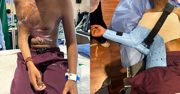
NPWT prepares larger at-risk wounds for delayed closure. It does not lead to epithelialisation. Wounds treated with NPWT can be closed primarily, grafted closed, or allowed to granulate and close with scar contraction [Figure 3].
Conclusion
NPWT has expanded to many types of wounds from complex traumatic wounds to prevention of post-operative infection. A review of the evidence of various forms of NPWT therapy can be found in the paper by the Greater Bay Areas Chronic Wound Care Alliance (2026).