Epidermolysis bullosa (EB) is a genetic disorder caused by mutations in a small number of genes important for skin function and structure (Prasad, 2011). It is a chronic, incurable, multisystemic and heterogenous genetic disorder characterised by skin fragility, resulting in blistering from minimal trauma or occurring spontaneously (Has et al, 2020; Martins Freitas et al, 2025).
The exact prevalence of EB is unknown, but it has been estimated that worldwide, EB affects approximately 1 in 17,000-20,000 live births, with 500,000 cases estimated globally (Featherstone, 2007; Prasad, 2011; Mellerio et al, 2023). EB incidence and prevalence vary across countries (Has et al, 2023). A collation of available evidence cites an overall EB prevalence (per million) of 11.1 in the US, 4.03-5.16 in Japan, 34.8 in England and Wales and 54.03 in Germany (Has et al, 2023).
EB is categorised into four groups according to the level of skin cleavage and the underlying genetic mutation [Table 1]: EB simplex (EBS), junctional EB (JEB), dystrophic EB (DEB) and Kindler syndrome (KS; Has et al, 2020). EBS is generally the mildest and most common form of EB (accounting for 40% to 70% of cases), while JEB (5% to 20% of cases) and DEB (25% to 50% of cases) are less common and can affect patients more severely (Has et al, 2020; Mellerio et al, 2023). KS is very rare (<1% of cases; Mellerio et al, 2023). More than 30 different subtypes of EB are currently recognised (Has et al, 2020; Khanna and Bardhan, 2024; Martins Freitas et al, 2025).
EB is characterised by skin fragility that arises secondary to structural defects in the dermo-epidermal junction (Khanna and Bardhan, 2024; Nyström, 2025). The type of EB is determined by the level of the skin at which separation of skin layers occurs (Snelson, 2020). As a result of this fragility, the skin becomes susceptible to damage from mechanical stress, minor trauma and shear forces, leading to blistering, erosions and ulceration (Alexandru et al, 2025). Severe instances of EB result in a spectrum of symptoms ranging from a few local blisters to more generalised skin and mucosal blistering and wounding. Consequently, patients with some forms of EB are at greater risk of infection and disabling deformities (e.g. joint contracture) due to severe scar formation (Tartaglia et al, 2021; Jayawardena et al, 2024). These patients may also develop aggressive cutaneous malignancies (e.g. squamous cell carcinoma), leading to early mortality (Montaudié et al, 2016; Prodinger et al, 2019). The presence of blisters and open wounds is accompanied by severe pruritus and pain, symptoms related to both irritation of damaged skin and inflammatory processes (Niebergall-Roth et al, 2024; Papanikolaou et al, 2021). EB guidelines recommend that “every effort should be made to treat the intense pruritus” to minimise scratching that can lead to further skin damage (Pillay and Clapham, 2018).
To date, there are no approved therapies for EB. An important aspect of EB care is a multidisciplinary approach, combining wound care with pain and itch management, provided through the use of appropriate dressings, pain control and management of secondary infections, which can worsen the patient’s condition (Denyer et al, 2017; Has et al, 2021; Wang et al, 2025). Wound management is challenging due to skin fragility, and a key principle of EB lesion management is the use of atraumatic dressings to prevent new blistering, minimise damage to periwound skin, and reduce pain during removal (Denyer et al, 2017; Pillay and Clapham, 2018; Danescu et al, 2024). Pruritus is one of the most common symptoms across all EB subtypes (Papanikolaou et al, 2021). It occurs at blistered or wounded sites or manifests as a generalised phenomenon, affecting both intact skin and healing wounds. Scratching in EB can irritate and tear already fragile skin, worsen existing wounds and create new blisters, with the so-called ‘itch-scratch-blister cycle’ contributing to chronic wounding (Danial et al, 2015; Mellerio et al, 2023). Skin inflammation secondary to barrier disruption, wound healing cascades and dysregulated activation of epidermal sensory nerve endings are likely involved in the pathophysiology of pruritus at both molecular and cellular levels (Papanikolaou et al, 2021).
The blisters that occur with EB may arise in response to minor trauma or friction, or develop spontaneously due to underlying inflammatory processes. Their development is directly related to alterations in structural components of the skin, such as collagen and keratin, which, when compromised, cause separation of the epidermal layers and lead to lesion formation. Intense itching is a common symptom, triggered both by inflammatory responses and by the healing of lesions (Bchetnia et al, 2024).
Effective wound care is imperative in the management of EB and includes strategies such as gentle cleaning, use of non-adherent dressings, protection from further trauma, infection control and palliative management of complications (El Hachem et al, 2014; Pillay and Clapham, 2018; Has et al, 2021; Danescu et al, 2024). Hydrogel dressings are an effective option for managing EB lesions and wounds by providing a moist healing environment, protecting the skin, supporting autolytic debridement, and alleviating pruritus (Denyer et al, 2017).
HydroTac® Transparent (PAUL HARTMANN AG), a single-use sterile transparent hydrogel dressing composed of hydrated polyurethane polymer and glycerin, offers a treatment option to address the challenges faced by healthcare professionals managing EB wounds. It provides itch relief, a cooling effect and maintains a hydroactive environment (Zoellner et al, 2007). The dressing’s low adherence enhances comfort, facilitates autolytic wound debridement, supports an optimal moist healing environment and protects damaged skin from friction and further trauma, while also reducing pain (Zoellner et al, 2007; Souter and Edwards, 2010; Klode et al, 2011; Bazire et al, 2015). EB is a representative example of a group of skin disorders characterised by skin fragility, comprising several classical types (Has et al, 2020).
The primary objective of this case series was to evaluate the efficacy of the hydrogel dressing in controlling pruritus, with secondary objectives of enhancing cutaneous hydration, modulating local inflammatory responses and reducing eschar in lesions of patients with DEB.
Materials and methods
A case series evaluation was undertaken on patients with DEB. This study was a multi-centre, retrospective, non-comparative case series investigation evaluating the use of HydroTac® Transparent hydrogel dressing in patients with DEB. The study centres were: the Instituto Médico FNakamura, São Paulo, Brazil (n=4) and an outpatient clinic in Germany (n=1).
Ethical approval and patient consent
This study received ethical approval from the Institutional Review Board (Number 8272500, Certificado de Apresentação para Apreciação Ética (CAAE) Number 95101925600000196). Written informed consent was obtained from all patients, including for the use of photographs. Written informed consent was obtained from the parent(s)/guardian(s) of paediatric patients.
Study dressing
HydroTac® Transparent is a single-use sterile transparent hydrogel dressing consisting of hydrated polyurethane polymer and glycerin with an outer semi-permeable layer to prevent bacterial penetration. The dressing is able to release moisture and absorb a limited amount of wound exudate (Alexander-Sinclair et al, 2025). HydroTac® Transparent allows for atraumatic dressing changes and its transparent properties allow wound inspection without removal of the dressing. Additional fixation is required using a suitable secondary dressing.
Treatment protocol
The treatment duration was determined based on the disease state and clinical assessment. Inclusion criteria were patients with a clinical diagnosis of DEB, the presence of intense pruritus, the presence of exudative lesions and agreement to participate in the study.
All participants were previously evaluated by a dermatologist or specialised wound care nurse, who recorded the location and characteristics of the skin lesions, including the presence of blisters, exudation, erythema and risk of infection. In Brazil, monitoring was carried out by a dermatology nurse specialised in wound care and dressings were changed every 48 hours or daily, depending on the level of exudate and the skin sensitivity presented by each patient.
Paediatric patients were monitored for a period of 30 days, with medical reassessments at the beginning and end of the observation period. Adult patients were monitored for 15 days, with the use of the dressing suspended after the remission of clinical symptoms (especially pruritus).
Intervention and assessment
All participants were initially evaluated and monitored throughout the study by a dermatologist/wound specialist nurse who recorded baseline characteristics and documented the response of skin lesions, including the presence of blisters, level of exudation, erythema, and risk of infection.
In Brazil, HydroTac® Transparent was changed every 48 hours or daily, depending on the level of exudate. A conforming bandage with latex-free cohesive coating (Peha-haft®) was used to ensure adequate fixation of the study dressing. Paediatric patients were monitored for up to 30 days and adult patients were monitored for 15 days. In the case of the German patient, HydroTac® Transparent was applied to the back to cover exuding eschar for 5 hours. Fixation was with Peha-haft® elastic bandage (PAUL HARTMANN AG, Germany).
Assessment included:
- Wound location.
- Wound characteristics (blisters, open or closed, exudate, eschar, etc).
- Pruritus (assessed on a scale of intensity, 0–10, where 0 indicates no itching).
- Cutaneous hydration (assessed by skin pinch test).
- Signs of inflammation (assessed by visual inspection).
- Presence of slough/eschar (assessed by visual inspection).
Results
In this case series, five patients (three male, two female) with DEB lesions were included. The patients’ lesions were treated using HydroTac® Transparent hydrogel dressing.
Case 1
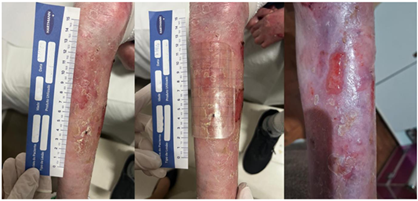
A 10-year-old boy presented with DEB lesions on the dorsum of the right foot. HydroTac® Transparent was applied to the EB lesions and was changed every 48 hours, and progress was monitored for 30 days. After commencement of treatment with HydroTac® Transparent, lesions showed significant improvement in skin hydration, clearer delimitation of the blister and visible reduction in the inflammatory process. There was also a reduction in itching, initially rated as intensity 9 (on a scale of 0 to 10), reducing to intensity 0 during the use of the dressing. No new blisters developed while using the dressing and existing blisters did not exhibit any signs of worsening. Removal of the dressing was considered atraumatic, without compromising the integrity of the adjacent skin. A gradual decline in associated pain was observed during the follow-up period. [Figure 1]
Case 2
A 15-year-old male presented with DEB lesions on the lower limb. HydroTac® Transparent was applied to the EB lesions and was replaced every 48 hours, with progress monitored over a 30-day period. After commencement of treatment with HydroTac® Transparent, lesions showed improvement in skin hydration, clearer delimitation of the blister and visible reduction in the inflammatory process. There was also a reduction in itching (from an intensity of 9, reducing to intensity 0 during the use of the dressing). No new blisters developed while using the dressing and pre-existing blisters did not worsen. Associated pain also showed a progressive reduction throughout the follow-up. The removal of the dressing was deemed atraumatic and did not compromise the integrity of the surrounding skin. [Figure 2]
Case 3
A 39-year-old woman with DEB presented with a lesion on the knee region of the left leg. The patient had a history of chronic pruritus resistant to conventional therapeutic approaches. HydroTac® Transparent was applied to the EB lesions, with dressing change periods of up to 48 hours and follow-up conducted for 15 days. HydroTac® Transparent was fixed with the aid of a cohesive bandage (Peha-haft®), and when required, supplemented with the use of tubular meshes already used by the patient in their daily routine. After starting treatment with HydroTac® Transparent, visual inspection indicated that there was an improvement in the general appearance of the skin in the knee region, with visibly increased hydration and complete relief from itching during the period of use of HydroTac® Transparent. Pruritus, initially of intensity 9, reduced to intensity 0 with the dressing in place. The alleviation of itching did not persist in the absence of the dressing and the pruritus recurred upon discontinuation of HydroTac® Transparent. The patient reported that it was difficult to apply the dressing to the joint area, but this did not affect the correct usage of the dressing. It was noted that, in this patient, the use of the dressing did not prevent new blisters from forming. [Figure 3]
Case 4
A 62-year-old woman with DEB presented with lesions in the left and right armpit. There was a history of chronic pruritus resistant to conventional therapeutic approaches. HydroTac® Transparent was applied to the EB lesion, with the dressing remaining in place for up to 48 hours. Follow-up was conducted for 15 days. HydroTac® Transparent was secured using a cohesive bandage (Peha-haft) and, in some instances, supplemented with tubular meshes already used by the patient in their daily routine. After treatment with HydroTac® Transparent, there was an improvement in skin hydration and significant relief from itching. Pruritus, initially of intensity 9, was completely controlled while the dressing was in place, reducing to intensity 0. The improvement in itching did not persist without the use of the dressing and itching returned upon discontinuation of HydroTac® Transparent. The dressing remained in place for 48 hours and there was an observable reduction in the size of the lesion. [Figure 4]
Case 5
A 29-year-old man presented with DEB lesions on his back; 75% scarring, 15% open wounds, and 10% crust. The periwound skin was dry with some necrotic tissue/eschar. The hydrogel dressing was applied to the EB lesions where eschar was observable on the back and, for fixation, Peha-haft was used to ensure adequate fixation. The patient independently performed wound care with his mother aiding placement of the dressing in difficult-to-reach areas. Due to the transparent nature of HydroTac® Transparent, the patient was able to monitor the wound and recognise when the dressing required changing (as the dressing becomes opaque due to exudate). After 5 hours of application, there was a visible reduction in wound eschar, with an associated increase in a clean, granulating wound bed. Once the eschar was removed, there was a reduction in itching experienced by the patient, with no pruritus in the area where the dressing was applied. The patient experienced no pain upon removal and no new blistering was observed. The patient reported an improvement in skin mobility. [Figure 5]
Summary
The study demonstrated improvements in DEB symptom management across all five patients (paediatric and adult), particularly for controlling pruritus, reducing the inflammatory status of lesions or wound tissues, improving skin hydration and facilitating the debridement of eschar. Most patients (4/5) noted improvements in the general appearance of the skin. Pre-existing lesions did not worsen over the course of the observation period and, although the formation of new blisters was not prevented, clinicians noted that these lesions appeared to develop less frequently than expected based on clinical experience. Removal of dressings at dressing change was atraumatic and there was no evidence of dressing-related trauma to perilesional skin.
Discussion
Pruritus is a major challenge in the management of EB, often more distressing than pain and linked to insomnia and depression (Danial et al, 2015; Denyer et al, 2017). It affects both intact skin and healing wounds across all EB subtypes and can begin at birth, significantly impairing quality of life (QoL) and contributing to depression, anxiety and social isolation (Papanikolaou et al, 2021). The itch-scratch cycle exacerbates skin damage, leading to blistering, delayed healing and increased infection risk. Intense pruritus is also considered part of the broader pain spectrum and inflammatory pathways are implicated in its pathogenesis (Metz and Ständer, 2010; Denyer et al, 2017; Bchetnia et al, 2024). Hydrogels (including hydrogel dressings), known for their cooling effect, have been used to lower wound temperature in burns and to relieve itch in EB (Denyer et al, 2017; Surowiecka et al, 2022). Cooling is a recognised method of reducing itch and pain (Palkar et al, 2018). Patients (paediatric and adult) with DEB receiving HydroTac® Transparent reported reduced pruritus, resulting in improved comfort and patient well-being, though the antipruritic effect is temporary and limited to the duration of dressing application. HydroTac® Transparent provides a soothing and cooling effect to wounds and this cooling effect may aid in the reduction of itching (Bazire et al, 2015; Palkar et al, 2018), suggesting that this dressing may be particularly beneficial for patients who live in countries such as Brazil with higher ambient temperatures.
Various inflammatory signalling pathways are involved in the mechanisms underlying EB pathology (Bchetnia et al, 2024). HydroTac® Transparent appeared to reduce local inflammation, as evidenced by decreased lesion inflammation and a lower incidence of blister formation (as noted by clinicians). Pain is associated with inflammation and has been shown to have detrimental psychological effects on patients as well as on healing progression (Woo, 2008, 2012; Matsuda et al, 2019). Clinicians noted a reduction in pain over the course of the observation study, suggesting that HydroTac® Transparent may play a role in modulating inflammation. More direct evidence is needed from further, larger-scale studies. A reduction in pain by HydroTac® Transparent contributes further to improved EB patient comfort and patient well-being.
For individuals with EB, wound dressings must be atraumatic (minimising pain and further skin damage) and low adherence (reducing pain and skin stripping during dressing changes; Denyer et al, 2017). This is crucial due to the extreme fragility of their skin, which blisters and tears easily with even minor friction or pressure. Clinical experience suggests that the ‘ideal dressing’ for an EB wound includes being non-adherent and atraumatic (Ly and Su, 2008). The results demonstrated the ease of application and removal of the dressing, without causing additional trauma to the skin, which was of paramount importance in this patient group.
Limitations
The main limitation of this study is that the populations analysed are small and studies with larger data sets from more patients would provide a basis for further developing evidence-based medicine. Being a rare disease with a low life expectancy, randomised controlled trials are difficult. Therefore, studies with larger populations are encouraged to confirm the effectiveness of HydroTac® Transparent and support its use in the treatment and management of EB. Longer follow-up periods will help to better evaluate the impact of the dressing on sustained outcomes.
Conclusion
This small observational study supports the use of HydroTac® Transparent as an integral part of the care protocol for patients with DEB, particularly for the control of inflammatory symptoms and pruritus. The results from this clinical case series have demonstrated that HydroTac® Transparent can be successfully used as part of comprehensive, symptomatic care for both paediatric and adult patients with DEB.
Additional studies with a larger number of participants and prolonged follow-up may further expand understanding of the therapeutic role of this type of dressing group, contributing to the development of evidence-based approaches to the care of EB.