Stage IV heel pressure ulcers with exposed bone and underlying osteomyelitis represent one of the most challenging scenarios in limb preservation, particularly among elderly patients with multiple comorbidities. The posterior calcaneus is uniquely vulnerable due to limited soft tissue coverage, high mechanical loading, and compromised perfusion, making reconstruction and healing especially difficult in medically complex individuals. When ulceration progresses to stage IV disease, the risks of deep infection, prolonged morbidity, and major amputation increase substantially (Thomas, 2001; Brem and Lyder, 2004; Miller, 2013).
Negative pressure wound therapy (NPWT) has become a cornerstone modality in the management of complex lower-extremity wounds, promoting granulation tissue formation, reducing oedema, managing exudate and stabilising the wound environment (Argenta and Morykwas, 1997; Morykwas et al, 1997).
Traditional NPWT protocols typically employ pressures near 125 mmHg with dressing changes every 48–72 hours (Morykwas et al, 1997; Armstrong and Lavery, 2005). However, frequent dressing changes may disrupt fragile granulation tissue, increase patient discomfort and impose significant logistical burdens, particularly in rural settings where home health resources are limited (Borgquist et al, 2009; Rayner et al, 2009).
These challenges have prompted growing interest in pragmatic NPWT strategies that reduce treatment burden while maintaining clinical effectiveness, including lower-pressure settings and extended dressing intervals (Borgquist et al, 2009; Rayner et al, 2009).
This report describes the successful limb salvage of a complex stage IV heel ulcer in a 91‑year‑old man with diabetes, peripheral vascular disease, chronic oedema and post‑polio limb weakness, complicated by calcaneal osteomyelitis. Intraoperative bone cultures unexpectedly grew Candida glabrata, and the patient was also found to have fungaemia, necessitating systemic antifungal therapy with caspofungin. Surgical management included partial calcanectomy followed by localised antimicrobial augmentation using Stimulan (Biocomposites), a resorbable calcium‑sulfate antibiotic carrier mixed with amphotericin B, gentamicin and vancomycin. Biologic reconstruction was performed using a combination of amniotic allografts and decellularised dermal matrix to provide layered biologic coverage and structural support.
Adjunctive wound care incorporated additional advanced biologic materials and specialised interface dressings.
NPWT was delivered using a low‑resistance wound contact layer engineered to reduce the compressive forces transmitted to the wound bed during suction. Traditional reticulated open‑cell foam (ROCF) exhibits increasing resistance to flow as it collapses under negative pressure, requiring higher suction levels (≈−125 mmHg) to achieve adequate exudate evacuation, thereby generating substantial reciprocal positive pressure against the wound surface (Kairinos et al, 2009a, 2009b). This compression has been shown to impair microvascular perfusion in a dose‑dependent manner, with perfusion decreasing progressively as suction increases (Kairinos et al, 2009a, 2009b; Borgquist et al, 2011). This interface dressing maintains open microchannels that permit efficient fluid evacuation at substantially lower suction levels, reducing the need for high negative pressures and mitigating the perfusion‑limiting effects described in foundational physiologic studies (Kairinos et al, 2009a, 2009b; Borgquist et al, 2011; Biermann et al, 2020). By lowering interface resistance and shear, the system supports reduced‑pressure therapy (50 mmHg) and extended dressing intervals, allowing the dressing to remain undisturbed for 7–10 days without compromising seal integrity or exudate management. This enabled continuous NPWT over a 6‑week period in a rural outpatient setting where frequent dressing exchanges would have been impractical. The combined strategy, integrating staged surgical debridement, targeted local antimicrobial delivery, advanced biologic grafting, and reduced‑pressure extended‑interval NPWT, resulted in successful wound closure and avoidance of major amputation in a medically fragile, high‑risk patient.
Calcaneal osteomyelitis itself carries a particularly high risk of both limb loss and mortality. The heel is biomechanically and anatomically unforgiving, and once infection penetrates the calcaneus, the likelihood of successful reconstruction decreases substantially. Multiple studies have shown that patients with hindfoot osteomyelitis experience disproportionately high rates of below‑knee amputation (BKA), often exceeding those seen in forefoot or midfoot infections (Pinzur, 2004; Lipsky et al, 2020; García‑Morales et al, 2012).In many centers, BKA remains the default surgical recommendation due to concerns regarding persistent infection, prolonged wound burden, and limited soft‑tissue options for durable coverage (Lipsky et al, 2020; García‑Morales et al, 2012; Attinger and Brown, 2012).As a result, attempts at limb salvage in this population are frequently avoided, despite the profound functional and survival implications associated with major amputation (Thorud et al, 2016; Izumi et al, 2009). These realities underscore the need for pragmatic, resource‑sensitive strategies capable of achieving infection control and wound closure while preserving limb function in medically fragile patients.
Extended interval NPWT is particularly relevant in rural environments, where limited home health availability often constrains adherence to traditional 48–72 hour dressing protocols. Adapting NPWT delivery to real world resource constraints may expand limb salvage opportunities for high risk patients.
Case presentation
The patient was a 91‑year‑old man with extensive multimorbidity contributing to impaired wound healing and progressive tissue breakdown. His medical history included insulin‑dependent type 2 diabetes (HbA1c 7.2% at initial encounter), peripheral vascular disease, chronic bilateral lower‑extremity oedema, post‑polio limb muscle weakness, peripheral neuropathy, atrial fibrillation (long‑term apixaban therapy), chronic steroid use with adrenal insufficiency, lumbar spinal stenosis with gait instability, recurrent urinary tract infections requiring a suprapubic catheter, and a history of DVT/PE. Additional chronic conditions included hypothyroidism, hyperlipidaemia, osteoarthritis, osteoporosis, gastro-oesophageal reflux disease, depression and chronic pain syndromes. His medication regimen reflected this complexity, including basal insulin, glipizide, prednisone, torsemide, apixaban, oxybutynin, sertraline, pantoprazole and multiple topical agents.
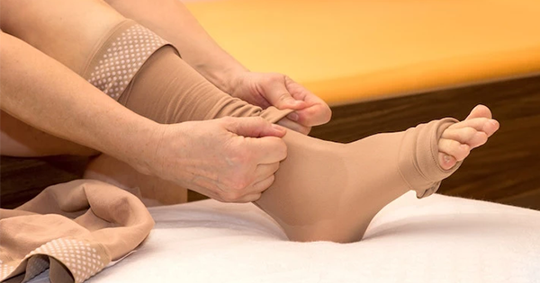
Functionally, the patient was wheelchair‑dependent, with limited ability to reposition independently due to bilateral leg weakness, left foot drop and chronic post‑polio deficits. He had no history of tobacco, alcohol or substance use. Prior to surgical referral, he had been using long‑standing compression garments, Velcro compression wraps, and shin guards to reduce trauma to his legs; however, his older compression stockings had lost elasticity and were no longer adequately controlling oedema. Offloading efforts included a heel‑elevation boot, placement of wedges under the calves and strict avoidance of weightbearing on the left heel. Despite these measures, progressive oedema, impaired mobility and neuropathy contributed to recurrent pressure loading of the posterior heel.
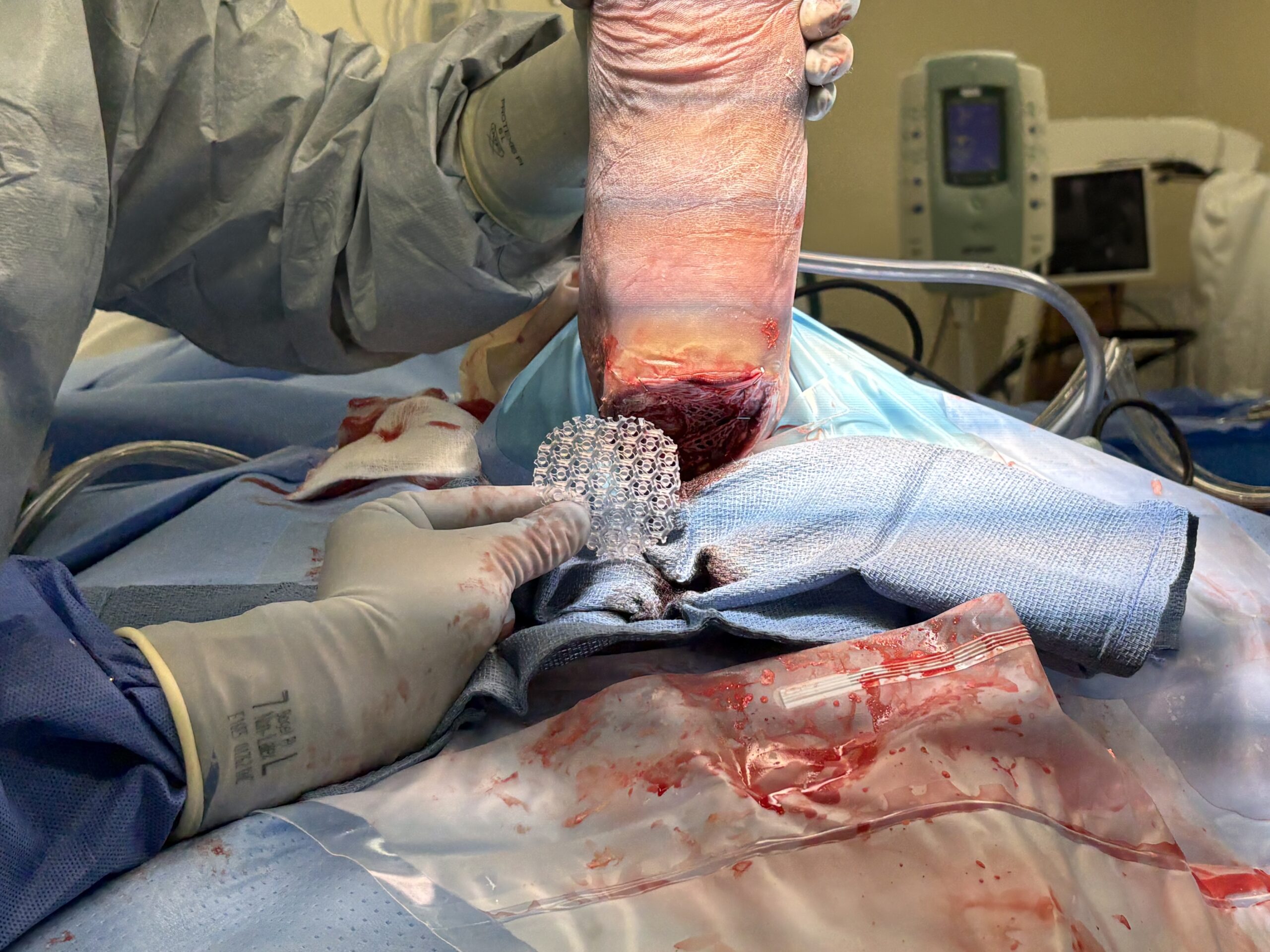
Pre‑surgical wound management included regular sharp debridement, saline irrigation, antimicrobial dressings, and attempts at oedema control. Pedal pulses were non‑palpable and the limb remained cool with significant pedal oedema. The heel ulcer continued to deepen, ultimately exposing bone. The patient’s metabolic control was acceptable, but not optimal for wound healing, and his chronic steroid use further impaired immune response. These factors, combined with limited mobility and persistent pressure despite offloading attempts, led to progression to stage IV ulceration with calcaneal osteomyelitis, necessitating operative intervention. On initial evaluation, the ulcer measured 9.5 × 7.5 × 0.9 cm (length × width × depth) with exposed calcaneal bone. Given the severity of infection and the high risk of proximal amputation, a limb salvage approach was pursued. A photo taken immediately after operative debridement and partial calcanectomy is shown in Figure 1.
The patient underwent a staged surgical approach to manage the advanced heel ulcer and underlying calcaneal osteomyelitis.
The first procedure consisted of operative debridement with partial calcanectomy to remove infected bone and non-viable soft tissue. Intraoperative bone biopsies were obtained to guide antimicrobial therapy. Immediately following calcanectomy, NPWT therapy was applied using Prevent (ClearChoice Therapeutics) as an interface dressing. NPWT was initially applied in an intermittent mode at 50 mmHg to assist with mobilisation of postoperative clotted blood and exudate into the canister.
Bone cultures were positive for C glabrata, an uncommon, but clinically significant, pathogen in calcaneal osteomyelitis. Fungal osteomyelitis is rare, but increasingly recognised in medically complex or immunocompromised patients, often requiring prolonged systemic antifungal therapy in addition to surgical source control. The patient was also found to have fungaemia, underscoring the severity of systemic involvement. After an infectious disease consultation, systemic antifungal therapy with caspofungin was initiated.
Once bone culture results were available, the patient returned to the operating room 4 days after the initial procedure for definitive local antimicrobial augmentation and biologic reconstruction. Multiple drill holes were created within the residual calcaneus using a 0.45 cm drill, and these channels were filled with Stimulan mixed with amphotericin B, gentamicin and vancomycin, and injected into each drill hole prior to polymerisation, allowing the material to fully conform to the geometry of the prepared channels and cure in situ as a stable, high‑concentration antimicrobial depot. Following local antibiotic delivery, layered biologic coverage was applied using Matrion (LifeNet Health), an amniotic allograft providing growth factors and anti‑inflammatory properties, together with DermACELL AWM (LifeNet Health), a decellularised dermal matrix that supports structural reinforcement and tissue regeneration. This combination provided both antimicrobial control and biologic scaffolding to optimise healing over the exposed calcaneus.
NPWT therapy was applied again using Prevent as an interface dressing to stabilise the grafts, manage exudate, and promote granulation tissue formation at 50 mmHg on continuous suction, which was maintained for the remainder of treatment. An intraoperative photograph demonstrating the Prevent interface dressing being positioned over the biologic grafts during the second procedure is shown in Figure 2.
NPWT was maintained at a continuous pressure of 50 mmHg throughout the 6‑week treatment period, with the dressing left undisturbed for extended intervals of 7–10 days at a time. A reduced pressure of 50 mmHg was selected to minimise ischaemic risk in the posterior heel and to align with the Prevent interface design, which facilitates effective fluid management at lower pressures. The 7–10 day interval reflected both clinic scheduling constraints and the stability of the NPWT seal, allowing for consistent monitoring without disrupting granulation tissue. By week four, the wound demonstrated robust granulation tissue which had fully covered the calcaneus [Figure 3].
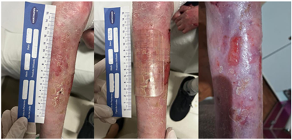
During these intervals, only the canister was exchanged as needed, allowing the interface dressing and seal to remain intact and minimising disruption to the wound bed. At regular clinic visits approximately one week apart (±3 days), the entire NPWT dressing was removed and replaced under direct supervision. These visits also allowed for comprehensive wound assessment, including local wound care, high‑resolution photographic documentation, and precise measurements of wound length, width, and depth.
Additionally, at each weekly clinic visit, following removal of the NPWT dressing and completion of wound assessment, an additional biologic support layer was applied to optimise healing conditions. Renograft (Surgenex) was used as an additional biologic graft layer to further support cellular integration, angiogenesis, and overall tissue regeneration and was placed directly onto the wound bed, followed by Supra SDRM (PolyMedics Innovations), a biodegradable matrix wound dressing designed to provide a temporary extracellular scaffold and support cellular migration, layered over the top.
To maintain stable positioning of the matrix and prevent shear during the subsequent NPWT cycle, the dressing was secured using Rylon‑1 (Bio Med Sciences), a single‑sided non-adherent wound interface that protects the underlying tissue while minimising disruption during removal. This adjunctive step was incorporated into every weekly dressing change to reinforce tissue regeneration and maintain a consistent biologic environment throughout the 6‑week treatment period.
A sequence of weekly clinic photographs demonstrating wound preparation, application of Renograft and Supra SDRM, placement of the Prevent interface, application of Rylon‑1 and NPWT film, and final NPWT device placement is shown in Figure 4.
This structured, extended‑interval protocol supported consistent monitoring while reducing cumulative tissue trauma and improving feasibility for outpatient management in a rural setting.
Clinical outcome
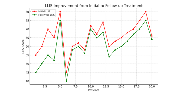
Over the 6-week course of NPWT, the wound demonstrated progressive improvement with healthy granulation tissue formation and reduction in wound depth. The extended-interval protocol allowed for stable wound management without evidence of excessive maceration, seal failure, or recurrent soft tissue infection. At 6 weeks, the wound had decreased to 2.0 × 2.3 × 0.1 cm, a significant improvement from the initial size of 9.5 × 7.5 × 0.9 cm (length × width × depth).
All measurements were obtained using a standardised disposable measuring guide and sterile depth probe to ensure consistency across visits. The wound showed a steady, quantifiable reduction in surface area and early resolution of depth, with the most rapid contraction occurring during the first 3 weeks of therapy.
Despite the presence of calcaneal osteomyelitis with C glabrata and associated fungaemia, the combination of surgical source control, systemic antifungal therapy, localised antimicrobial cement application, biologic grafting, and low-pressure NPWT resulted in successful limb salvage and eradication of infection. No complications occurred during the extended‑interval NPWT protocol.
Following discontinuation of NPWT, the wound continued to epithelialise. Ultimately, major amputation was avoided, a favourable outcome in an elderly patient with significant comorbidities and a high-risk stage IV heel ulcer.
Discussion
Stage IV heel pressure ulcers complicated by osteomyelitis remain among the most limb-threatening conditions in podiatric and wound-care practice. The heel’s limited soft tissue envelope, combined with mechanical pressure and vascular compromise, makes reconstruction and healing particularly challenging in elderly patients with diabetes and peripheral vascular disease.
This case demonstrates several clinically important and targeted considerations:
- The feasibility of reduced-pressure NPWT.
- The benefit of extended dressing intervals in minimising wound trauma.
- The importance of adaptable wound-care strategies in rural settings.
- The complexity of fungal osteomyelitis in limb salvage.
- A targeted technique of localised antimicrobial delivery within the calcaneus.
Traditional NPWT settings often use approximately 125 mmHg, based on early foundational studies demonstrating enhanced granulation tissue formation (Morykwas et al, 1997). However, lower-pressure protocols may offer advantages in select patients, including improved comfort and reduced risk of ischaemic compromise in vulnerable tissues (Kairinos et al, 2009a).
Standard NPWT dressing changes every 48–72 hours are commonly recommended (Armstrong and Lavery, 2005). Each dressing removal introduces mechanical disruption and potential loss of newly formed tissue (Borgquist et al, 2009). The ability to extend dressing intervals to 7 days or more may reduce cumulative wound trauma and improve feasibility in rural settings where home health access is limited (Rayner et al, 2009; Willy et al, 2017).
Standard management of calcaneal osteomyelitis often involves prolonged systemic antibiotics and, in many cases, early consideration of below‑knee amputation. This case illustrates that a structured, biologically supported NPWT protocol can offer a viable alternative even in the presence of fungal infection.
Access to home health services is often limited in rural communities. Patients requiring frequent NPWT changes may face delayed care, excessive travel burden or premature discontinuation of advanced therapy. In this case, the extended wear time facilitated by Prevent allowed for consistent outpatient management, bridging a common rural wound care gap and enabling limb salvage in a setting where standard protocols may have been impractical.
Although this case focuses primarily on clinical outcomes, the economic and access implications are also important, particularly in rural settings. The extended interval NPWT approach reduced the need for frequent home health visits, lowering overall care burden and making the treatment feasible in a region with limited wound care resources (Arcury et al, 2005).
When considered against the substantial costs associated with a below knee amputation, including operative care, hospitalisation, rehabilitation, prosthetic fitting, long term maintenance, and the well documented increase in mortality risk, limb salvage generally represents a more favorable cost value scenario for both patients and health systems (MacKenzie et al, 2004; Driver et al, 2010; Fortington et al, 2013).
All products used in this case are commercially available in the US, including in rural markets, and were selected based on accessibility within our practice environment. While pricing varies by institution, the components employed here are generally obtainable in lower resource settings, further supporting the translational relevance of this treatment model.
C glabrata osteomyelitis represents an unusual and challenging pathogen. Candida osteomyelitis is uncommon overall and often presents diagnostic and therapeutic complexity, frequently requiring combined surgical and systemic antifungal management (Gamaletsou et al, 2012; Kullberg and Arendrup, 2015; Pappas et al, 2016). Clinicians should remain aware that atypical organisms may occur in chronic pressure ulcers, particularly in elderly or immunocompromised patients.
An additional unique aspect of this case was the creation of drill holes within the residual calcaneus, which were then filled with resorbable antibiotic‑loaded cement. The cement was injected into each drill channel before polymerisation, allowing it to flow into and completely occupy the prepared voids. This ensured intimate contact with the surrounding bone surfaces and allowed the material to cure in place, forming a stable, conforming antimicrobial depot. By delivering amphotericin B, gentamicin and vancomycin directly into the cancellous bone, this technique provided high local antimicrobial concentrations at the site of infection following partial calcanectomy. While antibiotic cement is well established in orthopaedic infection management, its targeted use within calcaneal drill holes for limb salvage represents an innovative adjunct that may warrant further study.
Limitations
This case report reflects the inherent limitations of a multifaceted limb‑salvage strategy, in which several therapeutic components were applied in a coordinated sequence. Because the novel NPWT interface, the reduced‑pressure extended‑interval protocol, the layered biologic grafts, and the targeted drill‑hole antimicrobial cement were used concurrently, it is not possible to determine the relative contribution of any single modality to the successful outcome. The favourable clinical trajectory probably reflects the synergistic effect of these interventions rather than the isolated impact of one component.
Additionally, as a single‑patient experience, the findings may not be generalisable to all patients with calcaneal osteomyelitis, fungal infections, or severe comorbidity burdens. Further research is needed to clarify patient‑selection criteria, define safety parameters for reduced‑pressure extended‑interval NPWT, and determine which elements of this therapeutic combination are essential versus supportive. These limitations underscore the need for systematic evaluation of adaptable, resource‑sensitive wound‑care models in broader populations.
Conclusion
This case underscores the successful limb salvage of a stage IV heel pressure ulcer complicated by calcaneal osteomyelitis and C glabrata fungaemia through a coordinated, multidisciplinary strategy tailored to the realities of rural healthcare delivery. The use of low‑pressure NPWT at 50 mmHg with extended 7‑day dressing intervals, made feasible by the Prevent interface dressing, significantly reduced dressing‑related trauma and enabled reliable weekly outpatient management despite limited home‑health resources. When combined with partial calcanectomy, targeted local antimicrobial delivery via drill‑hole antibiotic cement, biologic grafting, and systemic antifungal therapy, this approach achieved durable wound closure and avoided major amputation in a medically fragile, high‑risk patient.
These findings highlight the potential value of reduced‑pressure, extended‑interval NPWT protocols as pragmatic, resource‑sensitive solutions for complex wounds, particularly in rural or underserved settings where traditional NPWT schedules may be impractical. Further investigation is warranted to better define patient selection, safety parameters, and long‑term outcomes associated with this adaptable treatment model.
Ethics
This case report did not require formal institutional ethical approval because it describes a single clinical case without experimental intervention. Written informed consent for publication of all clinical details and images was obtained from the patient, in accordance with journal guidelines and the principles of the Declaration of Helsinki.
Authors’ contributions: ACE was responsible for the conception of the case report, clinical management, data interpretation, and drafting and revising the manuscript. GM and BM assisted in the clinical care of the patient and contributed to the preparation and refinement of portions of the manuscript. All authors reviewed and approved the final version of the manuscript.